


Centers of Research Excellence (CORE)
CORE labs help advance significant research in specific areas by providing dedicated imaging systems as shared resources. Our CORE labs focus on:
Small Animal Imaging
The UMass Chan Medical School (UMass Chan) Small Animal Imaging CORE was established in early 2008 in order to serve the Medical School and the extramural research community by providing state-of-the-art small-animal imaging.
The imaging technology available at the Small Animal Imaging CORE facility enables tomographic imaging of animals from mice to rabbits and marmoset monkeys. It includes:
-
Bioscan NanoSPECT/CT camera for x-ray computerized tomography (CT) and single photon computerized tomography (SPECT)
-
Philips Medical System MOSAIC camera for positron emission computerized tomography (PET)
-
Two optical cameras, the IVIS-100 (Caliper) and the Pearl NIR Imager (Li-Cor) for detection of near infrared fluorophors
Dr. Mary Rusckowski, Director of he Small Animal Imaging Center, located in Room SA-107A.
Image-guided Surgery
Located within the New England Center for Stroke Research, this CORE facility offers UMass Chan investigators state-of-the-art image-guided intervention to accelerate discovery of clinically viable strategies for the delivery of therapeutic agents. Although the Center’s focus lies in cardiovascular imaging and intervention, previous work has included selective vascular drug delivery for cancer treatment and orthopedic device implantation.
The angiosurgical suite is dedicated to preclinical research and is home to:
- The latest flat-panel detector x-ray system available today (Allura FD20 R3, Philips Medical Systems, Best, the Netherlands), which offers both cardiac and vascular imaging packages, 3-dimensional reconstruction angiography (3DRA), X-per™ cone beam computed tomography, motor driven tilt table, and various software prototypes that are not yet available for clinical use
- Vascular analysis software with the ViewForum system
Matthew Gounis, PhD, Technical Director
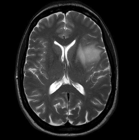 Advanced MRI Center
The Advanced MRI Center CORE facility provides the latest magnetic resonance imaging and spectroscopy capabilities to scientists, clinicians, government and industry, along with technical and clinical expertise for collaborative research.
The Center’s specialized techniques clarify functional, physiological and biochemical information from all organs of the body, enabling physicians to make more informed patient-care decisions. Additionally, researchers are better able to understand the mechanisms of such conditions as lung and heart disease, cancer, stroke, epilepsy, multiple sclerosis, lupus, rheumatoid arthritis, osteoarthritis, osteoporosis, back pain and injuries, autism, Alzheimer’s disease, bipolar disease and depression. This facilitates the development of new therapies that can be safely and continuously evaluated throughout patient treatment.
The Advanced MRI Center houses:
- 3T Philips Achieva whole-body scanner complete with a full array of clinical and animal RF coils
- Comprehensive physiologic monitoring system
- Anesthesia infusion pump
- Contrast injector
The Advanced MRI Center is on A-level of the UMass Chan Medical School.
Manojkumar Saranathan, PhD, Co-Director
Mohammed Salman Shazeeb, PhD, Co-Director
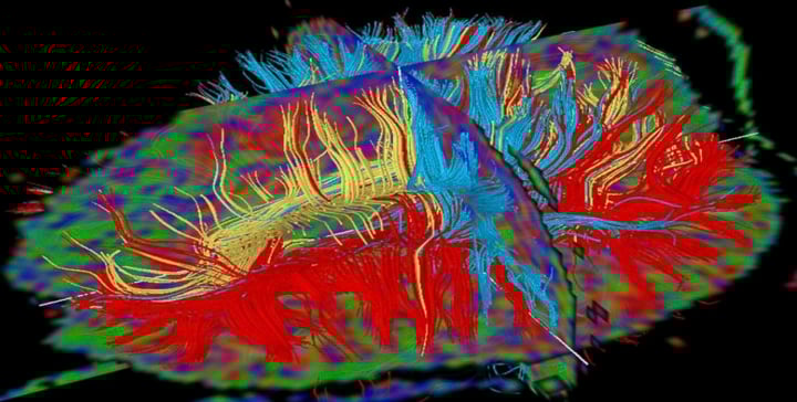 Image Processing and Analysis Core (iPAC)
The vision of the Imaging Processing and Analysis Core (iPAC) is to develop novel and existing imaging biomarkers to ameliorate disease conditions. iPAC provides image processing/analysis tools and services to help scientists and clinicians develop imaging biomarkers using different imaging modalities with the goal of:
- Detecting/diagnosing diseases to enable early treatment intervention;
- Tracking disease progression/regression to evaluate the medical condition, and;
- Monitoring treatment effects to facilitate therapeutic mechanisms.
Some of the services provided by iPAC include:
- Study design to identify useful imaging biomarkers;
- Perform modeling and simulations of imaging data;
- Implement imaging methods/protocols;
- Perform image processing/analysis from different imaging modalities including MRI, X-ray, CT, microCT, OCT, PET, SPECT, ultrasound, microscopy, etc., and;
- Prepare manuscripts/proposals in collaboration with investigators.
Mohammed Salman Shazeeb, PhD, Director
Matthew Gounis, PhD, Co-Director