CT Slices Templates for Skull Sculpture
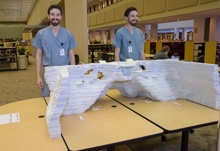
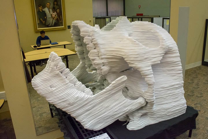
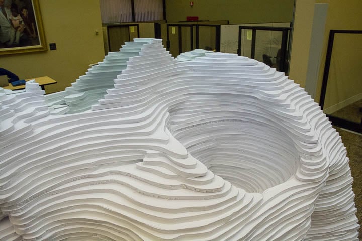
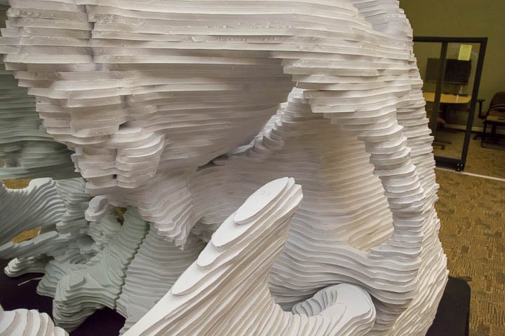
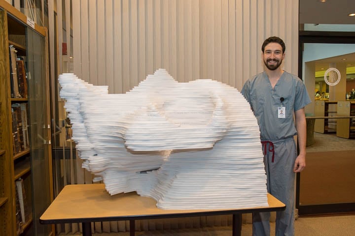 Fourth year Medical Student Matthew Breen created human skull model that currently resides in the UMass Chan Medical School Library. The hemisection sculpture depicts the left half of a human skull, which when rested against a mirror gives the illusion of a full skull model. It consists of approximately 270 pieces, in 107 “slices”. Each ½ inch thick slice represents 1 millimeter in real life. This translates to a roughly 13x scale model. The model will be upright when glued together. It will be 5.5 feet tall (66 inches cranio-caudal), 4.5 feet deep (53.5 inches antero-posterior) and 2.8 feet wide (34 inches left-to-right).
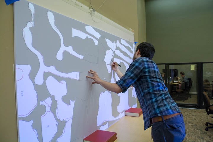
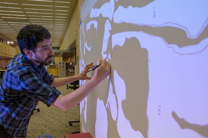
The pattern was generated by selecting a maxillofacial CT scan based on symmetry, lack of bony pathology and lack of dental hardware/artifacting. The scan was reformatted to 1mm slices (normally 1.5 in most scans). First all the CT images were batch cropped to only include the left side of the skull. Then each slice was placed on a separate Photoshop layer, and cut out with the pen tool. The outline of each layer was "stroked", or outlined, onto the layer below, so that when assembling the model it was clear where to place each piece. Each piece was then arranged onto a digital 8'x4' mock up of one of the foam boards to optimize space and materials. These were projected onto 8'x4'x1/2" foam boards. The outlines were traced in pen and the outline of the layer above in pencil so he knew where to cut and where to lay each piece.
The project took about 15 hours to trace the patterns digitally in Photoshop and 7 hours to trace the outlines on the foam boards. Matt estimates that it took him 50-60 hours to cut out the foam boards, a task which fortunately improved with practice. The cost of the materials was a little over $1300 including the mirror, foam board, glue and craft knives.
When asked what inspired him Matt said “Well, I've always loved sectional anatomy, and I thought this would be a really neat way of showing some of the intricate details of the skull. I polled the UMass Chan students to ask what topics in anatomy they found most difficult. Pelvis was number one, but head and neck was a close second. Many people were writing in that they found the pterygopalatine fossa especially difficult. I do hope to do a similar model of the pelvis, but I simply could not pass up the opportunity to make a sculpture of the most iconic piece of anatomy out there - the skull. I think it would be so cool to have a sort of "children's museum" for medical students one day.”
Matt is looking forward to a career in Radiology. He’s not sure what division, but says “neuro seems appropriate, doesn’t it?” He loves teaching and hopes to be involved in curriculum development for medical students or residents and to continue making artwork in his spare time.
According to Matt, the sculpture is currently quite fragile, and as tempting as it may be to sit in the sella turcica (Turkish saddle or seat), to please refrain from doing so. He also mentions that the foramina are completely patent – including the facial canal with genu and all! When it is finally glued together it will be pulled apart to reveal the sinuses, pterygopalatine fossa, and ear structures. All of the internal detail is present.
Matt acknowledges; “I'm very grateful to Dr. Max Rosen for his generous support for the project. Most of the materials cost including the mirror and foam board are thanks to Max with the help of Ella Covello and Jean Vigliotti in Radiology Academic Administration. I'd also like to thank Anne Gilroy for contributing her anatomical expertise and for her strong enthusiasm for the project. She has been a wonderful mentor through the process and will be greatly missed here at UMass. I'd also like to thank Dr. Peter Grigg, my capstone mentor who inspired me with hand-made visual aides in his lectures. Big thanks also to Dr. Andrew Chen in Neuroradiology for taking the time to help me through the process of locating a scan, and to Charlene Baron for helping me document the project as it unfolds. Lastly but certainly not least, thanks to Tess Grinoch and the staff of the Lamar Soutter Library for their logistical help through all of this and for tolerating all the little bits of foam board everywhere."
 |
 |
 |
 |
 |
 |
Anteroposterior Oblique from UMass Chan Radiology Dept on Vimeo.
BreenSkull-Lateral from UMass Chan Radiology Dept on Vimeo.
BreenSkull-SuperiorOblique from UMass Chan Radiology Dept on Vimeo.
BreenSkull-InferiorOblique from UMass Chan Radiology Dept on Vimeo.
BreenSkull-Filming-4hoursTimeLapse from UMass Chan Radiology Dept on Vimeo.


