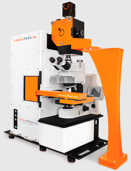
TissueFAXS SLQ Tissue Cytometer
High-Throughput, Multiplexed Imaging with the TissueFAXS SL
With the capacity to handle up to 120 slides per run, TissueFAXS SL is designed for efficient large-scale studies, whether you’re working with H&E, IHC, or highly multiplexed immunofluorescence assays. The system supports both widefield (up to 7-channel) and confocal fluorescence imaging (up to 6 channels), enabling detailed spatial analysis across a range of experimental needs.
Equipped with multiple objectives (from 2.5x overview to 40x oil immersion) and advanced detection using a Hamamatsu Orca Fusion BT camera, the platform delivers both panoramic tissue context and high-resolution cellular detail.
Importantly for spatial omics applications, TissueFAXS SL incorporates optimized filter sets for multiplex immunofluorescence, helping researchers capture complex marker panels with confidence and consistency.
Whether you’re mapping tissue architecture, validating spatial omics data, or building large imaging datasets, TissueFAXS SL bridges throughput, resolution, and multiplexing capability in a single, integrated platform.
Technical Specifications
Capacity: 120 slides
Imaging modes:
- Color camera (H&E, IHC, DAB)
- 7 channel fluorescence (widefield)
- 6 channel fluorescence (confocal, all colors listed below except 750nm).
Objectives: 2.5x (NA 0.085), 10x (NA 0.45), 20x (NA 0.5), 20x (NA 0.75), 40x oil (NA 1.3)
Cameras: PixeLINK (color), Hamamatsu Orca Fusion BT
Light source: Lumencor Spectra III
Spinning Disc: Crest V2
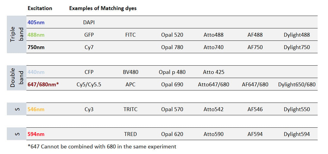
The TissueFAXS SL features Kromnigon Spectra Split filters optimized for multi-plexed IF staining. Compatible dye sets are listed in the table below. Please contact the SCOPE if you have questions about dye selection.