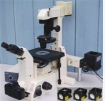
Nikon Diaphot 300 Wide Field Fluorescence
 Nikon Diaphot 300 Wide Field Fluorescence location: S5-213B status: in service reserve use
Inverted wide field fluorescence scope with color camera and DIC optics. Manual stage control.
This workhorse tissue culture microscope features advanced optical design for the shortest, most efficient light path. Incorporated as a part of the built-in turret assembly is a focusable Bertrand lens, photomask reticle and dark slide. The optical design utilizes a built-in binocular body, inclined at 45 degrees, and only one reflecting surface in the microscope body to reduce reflections and glare and maximize image contrast.
The stage is located slightly below the binocular head, permitting easy access to and viewing of the specimen area. The coaxial coarse and fine focusing system control the nosepiece, leaving the stage stationary and the specimen exposed for manipulation during observation.
Objective lenses:
DL 10X, Ph 1, n.a .0.3, dry
Plan Fluor 20X Ph 2, DLL n.a. 0.5, dry
Plan Fluor 40X Ph 4 DL n.a. 1.30 oil
Plan APO 60X, DIC n.a. 1.40 oil
Plan APO 100X, DIC n.a. 1.40 oil
Fluorescence filter cubes
UV-1A
This set, intended for applications using a mercury arc-discharge lamp, is designed to significantly reduce specimen autofluorescence and photobleaching by incorporating a very narrow bandwidth (10-nanometer) ultraviolet excitation cross section with a 40-nanometer gap between the dichromatic mirror and longpass emission filter.

Ultraviolet Excitation Filter Block UV-1A Specifications:
Excitation Filter Wavelengths: 360-370 nanometers (bandpass, 365 CWL)
Dichromatic Mirror Cut-on Wavelength: 380 nanometers (longpass, LP)
Barrier Filter Wavelengths: 420 nanometer cut-on (longpass, LP)
The UV-1A fluorescence filter combination is designed to minimize autofluorescence partially by utilizing only the i-line region of the mercury ultraviolet emission spectrum (centered at 365 nanometers). Consequently, this filter combination is recommended primarily for microscopes equipped with a mercury arc-discharge lamp. The longpass emission (barrier) filter used in the UV-1A combination is designed to collect signals at wavelengths exceeding 420 nanometers, enabling visualization of red, orange, yellow, green, and blue emission from fluorophores excited in the ultraviolet. The UV-1A filter combination is recommended when studying the following fluorophores: 7-amino-4-methylcoumarin-3-acetic acid (AMCA), anthroyl stearate, bisbenzamide, Calcein Blue, Cascade Blue, 4',6-diamidino-2-phenylindole (DAPI), FluoroGold, and Hoechst 33342/33258, among many others. The images presented in Figure 2 demonstrate the performance of this filter combination with a variety of ultraviolet absorbing fluorescence probes targeted at different intracellular locations.
-------------------------------------------------------------------------------------------------------------------------------------------------------
G-1B
The Nikon G-1B longpass emission filter set utilizes a narrow bandpass (10 nanometers) excitation filter, centered at 546 nanometers. It has increased separation between the cut-on wavelengths of the dichromatic mirror and emission filter (565 and 590 nanometers, respectively), enabling transmission of orange and red, with minimal yellow emission from fluorophores absorbing near the center of the green spectral region. Ultraviolet, visible, and near-infrared transmission spectral profiles for the Nikon G-1B filter combination are illustrated below in Figure 1.

Green Excitation Filter Block G-1B Specifications:
Excitation Filter Wavelengths: 541-551 nanometers (bandpass, 546 CWL)
Dichromatic Mirror Cut-on Wavelength: 565 nanometers (longpass, LP)
Barrier Filter Wavelengths: 590 nanometer cut-on (longpass, LP)
The G-1B filter combination is equipped with a narrow passband (10 nanometers) excitation filter, which minimizes autofluorescence. The center wavelength of the filter is positioned to correspond to the 546-nanometer emission line (referred to as the e-line) of a mercury arc-discharge source, which is the recommended application for the G-1B set. The longpass emission (barrier) filter used in this set is designed to collect fluorescence signals at wavelengths exceeding 590 nanometers, permitting visualization of orange and red emission, while blocking most yellow wavelengths.
The G-1B filter green excitation filter combination is recommended when investigating the following fluorophores: Alexa Fluors (532, 546, 555, 568, and 594), dichlorodimethoxyfluorescein (JOE), Alizarin Red, BODIPY probes, Calcium Orange, Cy3, Cy3.1.8, dioctadecyl tetramethylindocarbocyanine (DiI), ethidium bromide, FluoroRuby, hexachlorofluorescein (HEX), LDS 751-DNA, MitoTracker Orange and Red, R and B-phycoerythrin, POPO-3, PO-PRO-3, propidium iodide (PI), Pyronin B, RedoxSensor Red CC-1, RH probes (237, 414, 421, 795), many rhodamine derivatives, Sevron Brilliant Red, SYTO derivatives, SYTOX Orange, and Xylene Orange. The images presented in Figure 2 demonstrate the performance of this filter combination with a variety of green-absorbing fluorescence probes targeted at different intracellular locations.
--------------------------------------------------------------------------------------------------------------------------------------------
B-2E
This filter set is designed with a wide excitation bandpass (40 nanometers) in order to provide a relatively high signal-to-noise ratio with an adequate spectral absorption and excitation profile for most fluorophores responding to blue wavelengths. The bandpass emission filter has a center wavelength of 540 nanometers (40-nanometer bandpass), and in similarity to the other filter combinations in this group, the B-2E set employs a longpass dichromatic mirror, which has a cut-on wavelength of 505 nanometers.

Blue Excitation Filter Block B-2E Specifications:
Excitation Filter Wavelengths: 450-490 nanometers (bandpass, 470 CWL)
Dichromatic Mirror Cut-on Wavelength: 505 nanometers (longpass, LP)
Barrier Filter Wavelengths: 520-560 nanometers (bandpass, 540 CWL)
The B-2E filter combination is specifically designed with a wide excitation passband suitable for imaging specimens labeled with fluorescein isothiocyanate (FITC) and derivatives, in addition to providing sufficient excitation energy to enhance emission (relative to the B-1E set) from a range of fluorochromes with similar absorption characteristics. In order to exclude crossover from yellow, orange, and red wavelengths, the filter set incorporates a bandpass barrier filter that is identical to that included in the B-1E set, but with a longer bandpass center wavelength than that of the B-2E/C combination. Among the Nikon blue excitation filter sets, those in the E series, in general, produce images with a much higher signal-to-noise ratio than do combinations with longpass emission filters (the A filter category). In addition, the image background regions appear significantly darker with the bandpass filter combinations. Fluorescence emission occurring at longer wavelengths than green (the yellow, orange, and red spectral regions) is excluded from images collected using the B-2E filter combination (as illustrated in Figure 2).
The B-2E filter set is recommended when investigating the following fluorophores: Acridine Orange-DNA (excludes detection of Acridine Orange-RNA), Acridine Yellow, Alexa Fluor 488, BODIPY probes, Calcein, Calcium Green, carboxyfluorescein (FAM), Cy2, DiO probes, enhanced green and yellow fluorescent proteins (EGFP and EYFP), red-shifted green fluorescent protein (RsGFP), FITC, LysoTracker derivatives, MitoTracker Green, Oregon Green, Rhodamine 123 (and related xanthenes), Spectrum Green, SYTO probes, SYTOX Green, YO-PRO 1, and YOYO 1. The images presented in Figure 2 demonstrate the performance of this filter combination with a variety of blue light absorbing fluorescence probes targeted at different intracellular locations.
--------------------------------------------------------------------------------------------------------------------------------
Carl Zeiss AxioCam color camera. With a dynamic range of more than 1:2200, the highly sensitive2/3” CCD sensor acquires up to14 imagesper second in full resolution.
Documentation downloads: Nikon Diaphot manual.pdf