Does someone in your family have vitiligo? We are conducting a study to understand the causes of vitiligo and predict who might be at risk of developing it. We would like to invite siblings, children, or other close relatives of individuals with vitiligo who live in the United States to participate in this study.
Vitiligo is an autoimmune disease of the skin that causes white spots. It’s pretty distinctive and is frequently diagnosed by a dermatologist simply by looking at it, often with the aid of a special light called a Wood’s lamp. But I’ve seen patients in my Vitiligo Clinic, some who’ve traveled a very long distance (even other countries), who didn’t actually have vitiligo, but had diseases that looked like vitiligo. Sometimes I’ve had vitiligo patients who notice new spots on their bodies that they think are vitiligo, but they’re something else, often things that are even easier to treat. So, if you’re wondering if you have vitiligo but aren’t sure, maybe this blog will help you figure that out, and clarify what’s distinct to vitiligo versus its lookalikes!
There are different patterns of vitiligo
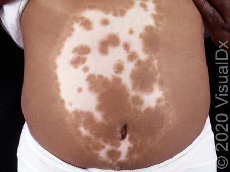
Vitiligo can sometimes have different appearances, so being familiar with the different patterns that can exist is helpful to recognize it and tell it apart from other conditions. For example, vitiligo can affect a small area (usually early on before it spreads), called focal vitiligo. It can become widespread, often called generalized vitiligo. It can affect just the lips and genitals, called mucosal vitiligo, or those areas plus the fingertips, called lip-tip vitiligo. It can affect the face and hands/feet, which is called acrofacial vitiligo. Universal vitiligo means that most of the skin has lost its pigment as vitiligo has spread all over, usually we reserve this term for when at least 80% is affected. Maybe most importantly, it can affect just one side of your body and stay in a small area, which is called segmental vitiligo. The details of these forms of vitiligo are for another blog post, but if you or your doctor knows these patterns it’s easier to determine if the spots are from vitiligo or a lookalike that causes the same pattern.
The physical exam and Wood’s lamp
The Wood’s lamp is a UVA light that looks dark purple, and it’s used by holding it close to the skin with all the lights in the room turned out. It’s the same light used for highlighting fluorescent colors in the dark, like “midnight bowling”, “fluorescent mini golf”, and clubs in the 70s. So be careful when going to one of these places that uses a “black light” if you have vitiligo, because your spots will glow! Maybe you’ll think this is cool, and want to find one. . .
When a patient has vitiligo, the Wood’s light makes all of the white spots fluoresce bright white, making them clearly visible in contrast to the normal skin color, even if the normal skin is very pale. It has something to do with proteins in the skin absorbing UVA light and then emitting (or shining back) a longer wavelength that’s in the visible spectrum, so your eyes can better see it. It’s an incredibly useful tool for a dermatologist and vitiligo specialist, because very few other diseases turn the skin white like this. So, when the spots glow under Wood’s lamp, it narrows down the possibilities by a lot. Combined with other pieces of information gathered by talking with the patient, we can usually diagnose vitiligo without a skin biopsy.
But what if it’s NOT vitiligo? What if the spots don’t fluoresce under Wood’s lamp illumination, or what if they do, but the other information doesn’t fit with what we know about vitiligo? Well, that’s when a dermatologist forms a “differential diagnosis”, or a list of diseases that might cause the skin changes. Next, we gather more information to narrow down this list to one or just a few possibilities, and maybe biopsy the skin to get a really close look under the microscope.
The first order of business is to determine, “Are these white spots actually depigmented, meaning they do not have ANY pigment in them?” So, to do this, I turn out all the lights in the room and turn on my Wood’s lamp. This is sometimes fun for my younger patients, because not just vitiligo glows under the light, but also certain colors of clothing, teeth, peanut butter left over from lunch, rubies (discovered they REALLY glow), and even lint. But I digress. I look for the white spots to enhance, or become more apparent, under the Wood’s lamp compared to room light, they kind of fluoresce. If this happens, the skin in the area is depigmented, meaning there is no pigment or melanin. This is always the case in vitiligo, and thus is an absolute requirement to diagnose it. If the spots do enhance, I know we’re on the right track.
Truly depigmented spots that are NOT vitiligo
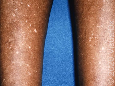
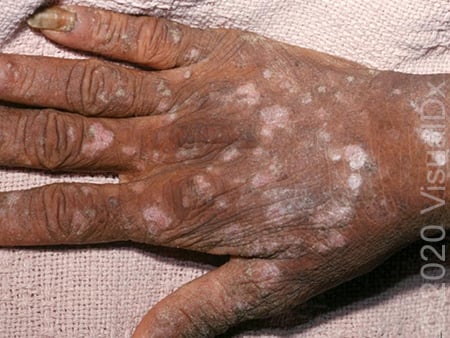
If the spots enhance under Wood’s lamp, they could be from vitiligo or a small number of other conditions. One is idiopathic guttate hypomelanosis (IGH), or small spots that appear on skin that has had chronic sun exposure (usually the shins and forearms, but sometimes the chest and back as well).
idiopathic guttate hypomelanosis (IGH)
Often these are hypopigmented, not depigmented (hence the name), but sometimes they are depigmented and enhance on Wood’s lamp. If they are evenly distributed (not clustered), remain small (usually 1-2mm in diameter but almost always under 5mm), and are on the right areas of the body, then they’re probably IGH and not vitiligo.
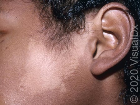
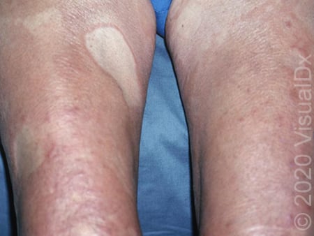
Another condition that rarely causes a true white spot is nevus depigmentosus, which is a birthmark that usually appears within the first few months of life, has jagged edges, usually doesn’t turn the hair white, and doesn’t grow in size like vitiligo – it may get larger as the child grows and the skin stretches, but not quickly the way vitiligo does (often within just a few months).
Nevus Depigmentosus
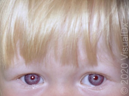
If your dermatologist isn’t sure if the spot is nevus depigmentosus or segmental vitiligo, a biopsy can tell for sure, but it’s not always necessary. I guess that albinism should be mentioned here, although that is present at birth and results in no skin color at all (would look like universal vitiligo), so it’s pretty easy to distinguish.
Albinism
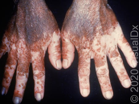
Finally, piebaldism causes depigmentation localized to just the front of the body (not the back), includes a “white forelock” or white hair at the front of the scalp, is present from birth, and usually runs in families. That’s about it for true white spots!
Piebaldism
Hypopigmented spots that are NOT vitiligo
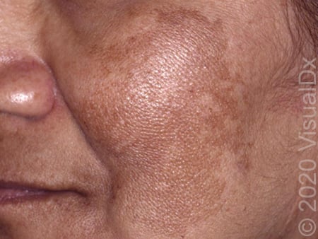
If the spots are not truly white, but hypopigmented and not depigmented (they don’t enhance by Wood’s lamp), then they are NOT vitiligo and could be any number of different diseases and conditions. I’ll list a few of the most common ones here. Both IGH and nevus depigmentosus (described above) can also be hypopigmented instead of depigmented. They look similar and are located on the same areas of the body. Nevus anemicus is a common birthmark that looks lighter than surrounding skin, although it is actually not different in pigment at all. It is due to a lower blood supply to that area of skin, so it is less pink than surrounding skin and thus looks lighter. It actually disappears completely for a few seconds when you apply pressure to the area because the surrounding blood in the vessels is pressed out. It reappears once the blood rushes back into it. Individuals with tuberous sclerosis can have light areas of skin called ash leaf spots, but they usually have other more cleare signs of this condition as well. Occasionally, darker skin from melasma can make it look like the normal skin is lighter, and thus could look a little like vitiligo.
Melasma
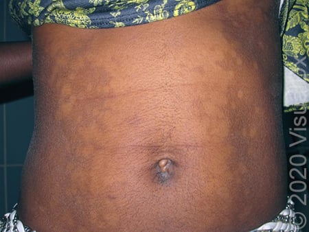
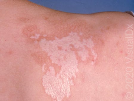
Tinea versicolor (also called pityriasis versicolor) causes lighter spots on the chest and back, get scaly if scratched with a fingernail, and are caused by a common fungus that isn’t dangerous. It’s easily treated with an antifungal therapy.
Tinea versicolor
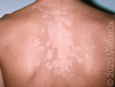
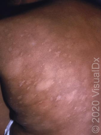
Progressive macular hypomelanosis looks a lot like tinea versicolor but without the scale, most commonly affecting the chest and back.
Progressive macular hypomelanosis
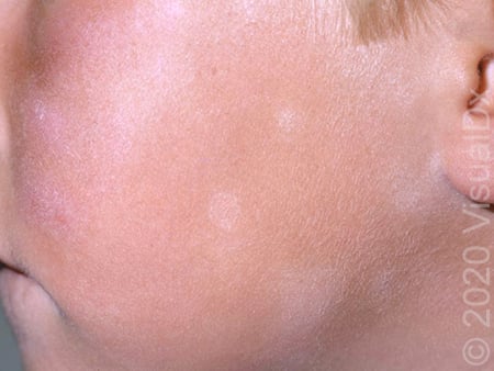
It is pretty easily treated with nbUVB therapy, similar to vitiligo. Inflammation or wounds can be lighter than the surrounding skin once they’re healed, this is called post-inflammatory hypopigmentation (PIH). Atopic dermatitis or eczema does this commonly on areas of the body where this disease occurs, such as the cheeks, front of the elbows, or back of the knees, and it’s sometimes called pityriasis alba when this is the case.
Pityriasis alba
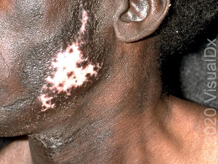
Discoid lupus (also called chronic cutaneous lupus) is usually located on the head and neck, causing lighter spots surrounded by dark areas, as well as permanent hair loss.
Discoid lupus
Lichen sclerosus et atrophicus (or just lichen sclerosus, LS) is usually located on the genitals and causes light spots (sometimes completely white). It usually also causes symptoms like itching or pain, with fissuring or open cracks in that area that can be sore. Intercourse can also be painful. This can cause long-term problems and should be treated aggressively.
Morphea causes hardening of the skin more than color change, although it can appear lighter than the surrounding skin. This should be treated to avoid permanent changes in the skin of that area, as well as prevent new spots from forming.
Morphea
Sarcoidosis can occasionally cause light spots on the skin that look like vitiligo, although it usually looks quite different. Sarcoidosis can also affect the lungs and other organs and should also be treated aggressively.
Hypopigmented mycosis fungoides (MF) or cutaneous T cell lymphoma (CTCL) is a very, very rare skin lymphoma that causes light spots on the skin most commonly only in sun-protected areas, or “bathing suit distribution”. This is not usually dangerous but should also be treated to prevent it from becoming a more aggressive form of the disease.
Hypopigmented mycosis fungoides
There are some VERY RARE infections that can cause light spots on the skin, and these include a form of leprosy (tuberculoid leprosy), secondary syphilis (a rash that occurs after the genital sore or chancre clears up), and pinta. Leprosy occurs most commonly in South Asia and South America (although interestingly it can be spread by armadillos in the southern United States) and is usually numb to light touch within the spot.
Leprosy
Syphilis is a sexually transmitted disease preceded by a genital sore (sometimes not noticed, though), and easily treated by penicillin once diagnosed.
Secondary syphilis
Pinta is only found in small areas of northern South America and causes white areas on the hands, wrists, and feet. I’ve never met anyone who has ever seen Pinta, so it’s not around much anymore.
Pinta
Your dermatologist can help!
Those aren’t ALL of the conditions that can cause white or light spots like vitiligo, but it’s most of them. Others are really rare and could require a lot of explanation better suited to residency training in dermatology, so I left them out! I don’t expect that this description will be sufficient to avoid a visit to a dermatologist because there’s a LOT more to diagnosing skin disease than knowing the names of diseases (however crazy they are) and a few lines of description, but it should help you wrap your mind around the possibilities and get some insight into how we’re thinking during a physical exam in our office. Remember, you could have BOTH vitiligo and another condition described here! It’s helpful to know which is which, so that your dermatologist can determine when and how to treat them. Now you can go to your dermatologist appointment armed with knowledge, which should help both of you figure out what your white or light spots are, and how best to treat them!
Does someone in your family have vitiligo? We are conducting a study to understand the causes of vitiligo and predict who might be at risk of developing it. We would like to invite siblings, children, or other close relatives of individuals with vitiligo who live in the United States to participate in this study.