Biologically, our mothers give us so much. Regardless of your relationship with your biological mother, they gave each of us vitally important biological gifts. In addition to approximately half of the DNA that makes up our genetic code, biological mothers also contribute most of the nutrients and other factors that help kick-start embryonic development.
The female germ cell, the egg or the oocyte, is about 10 MILLION times larger (by volume) than the male germ cell, the sperm1.
x
But what exactly does the oocyte carry in that large volume, and what does it do?
To learn more, we can turn to Dr. Sean Ryder in our department. Dr. Ryder’s fundamental scientific interest is understanding how cells in an embryo become the specialized organs and tissues found in an adult organism. Scientists refer to this process as “differentiation” or “cell fate specification,” in an effort to describe the complex changes that occur as a single cell that can literally become any other type of cell in the body divides to create other cells that make up the hundreds of different cell types in an adult human.
There are many factors that contribute to cell fate specification throughout the life of an organism, but Dr. Ryder’s focus is on the contents of that large oocyte cell. Specifically, Dr. Ryder is interested in the RNAs that can be found in oocytes. RNA is the molecule that acts as a transcript of the genetic material found in DNA that directs cells to make the proteins they need to survive and thrive. RNA from the oocyte is particularly important, since the early embryo isn’t immediately ready to start transcribing its own DNA. The RNA from the biological mother is therefore responsible for the first proteins made in a new embryo, and these proteins are essential for kick-starting the growth and development of the embryo. One of the most important things that starts happening when the cells of the embryo divide is differentiation, and the oocyte RNAs guide this process as well. Interestingly, turning off and getting rid of oocyte RNAs is also important for growth and differentiation as the embryo starts making its own RNA from its own brand new genome.
x
Some of you might be thinking – how can we possibly know all of this, since human single-cell embryos don’t just grow on trees (i.e. they are not abundant or accessible, nor can they be experimentally manipulated)?
x
Dr. Ryder and many other scientists work around the limitations of studying human embryos by instead studying embryos in “model” organisms, including microscopic worms that can produce hundreds of embryos in a few days. Even though worms and humans are very different, the challenge of cell fate specification has existed since multicellular organisms first evolved about 600 million years ago2.
x
The fundamental processes required to make cell types like muscle cells, neurons, and skin cells also developed millions of years ago, so a lot of these processes are remarkably similar between humans and worms.
An illustration of the nematode worm and common biological model system, C. elegans. The head of the worm is on the left, and several of its organs are labeled including the pharynx, intestine, gonad arm, spermatheca and uterus. Illustration by Leonora Martínez-Núñez, PhD.
Although we may share many developmental and cell fate specification processes with worms, the parts of our bodies that make oocytes or sperm (our reproductive systems) are very different. The species of worm used by scientists around the world (scientific name Caenorhabditis elegans) can reproduce by mating, with germ cells coming from two different worms (the way humans do it), or they can reproduce with germ cells coming from the same single worm (meaning the reproductive system of a single worm is making both oocytes and sperm). This is called hermaphroditic reproduction and is a helpful characteristic for scientists studying reproduction because all of the reproductive components are present in one single organism (any time you simplify a system, it makes it easier to study scientifically by reducing the number of variables at play). In worms, oocytes are made later in life, while sperm are made during a limited window of time before the worms become adults. The two meet and form an embryo that continues to develop for a while in the uterus of the hermaphrodite worm.
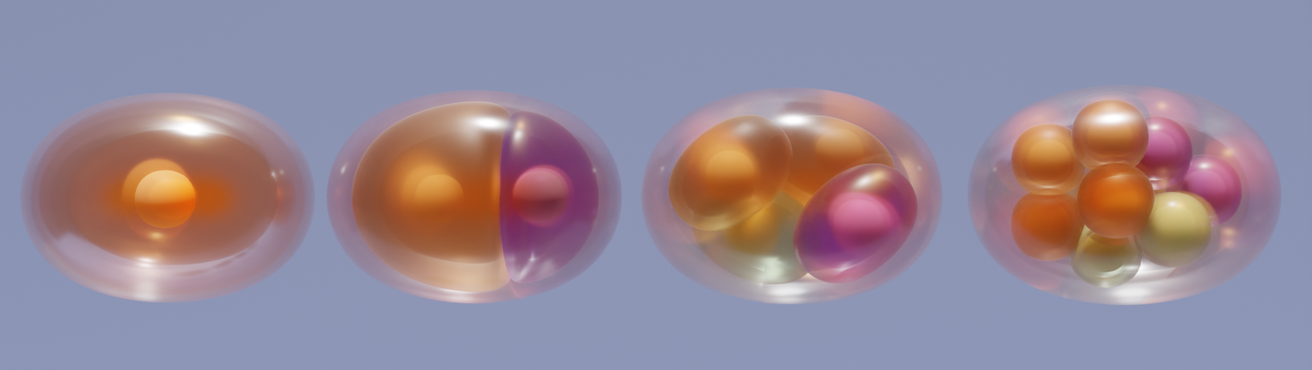
A figure depicting C. elegans hermaphroditic embryonic development. Female germ cell development begins in the stem cell niche maintained by the Distal Tip Cell (DTC). Early oocytes divide and then begin meiosis as they travel through the reproductive system. Fully-developed oocytes then come into contact with male germ cells (sperm) from the spermatheca, and then embryos develop in the uterus of the worm. The early stages of embryonic development are shown as well. Illustration by Leonora Martínez-Núñez, PhD. Figure adapted from Albarqi & Ryder, 2023.
An example of an important regulator of cell fate specification in the worm (and in humans) is the protein MEX-3, which was first described in worms in 1996. This protein is made from RNA produced in the female germline (oocytes) and helps to regulate differentiation in the early worm embryo. Specifically, this protein helps to ensure the proper differentiation of muscle cells. Despite all this knowledge, it still wasn’t clear how the mex-3 RNA was being controlled. After making mex-3 RNA, the oocyte has to find some way to keep it from being used to produce protein before the egg is fertilized.
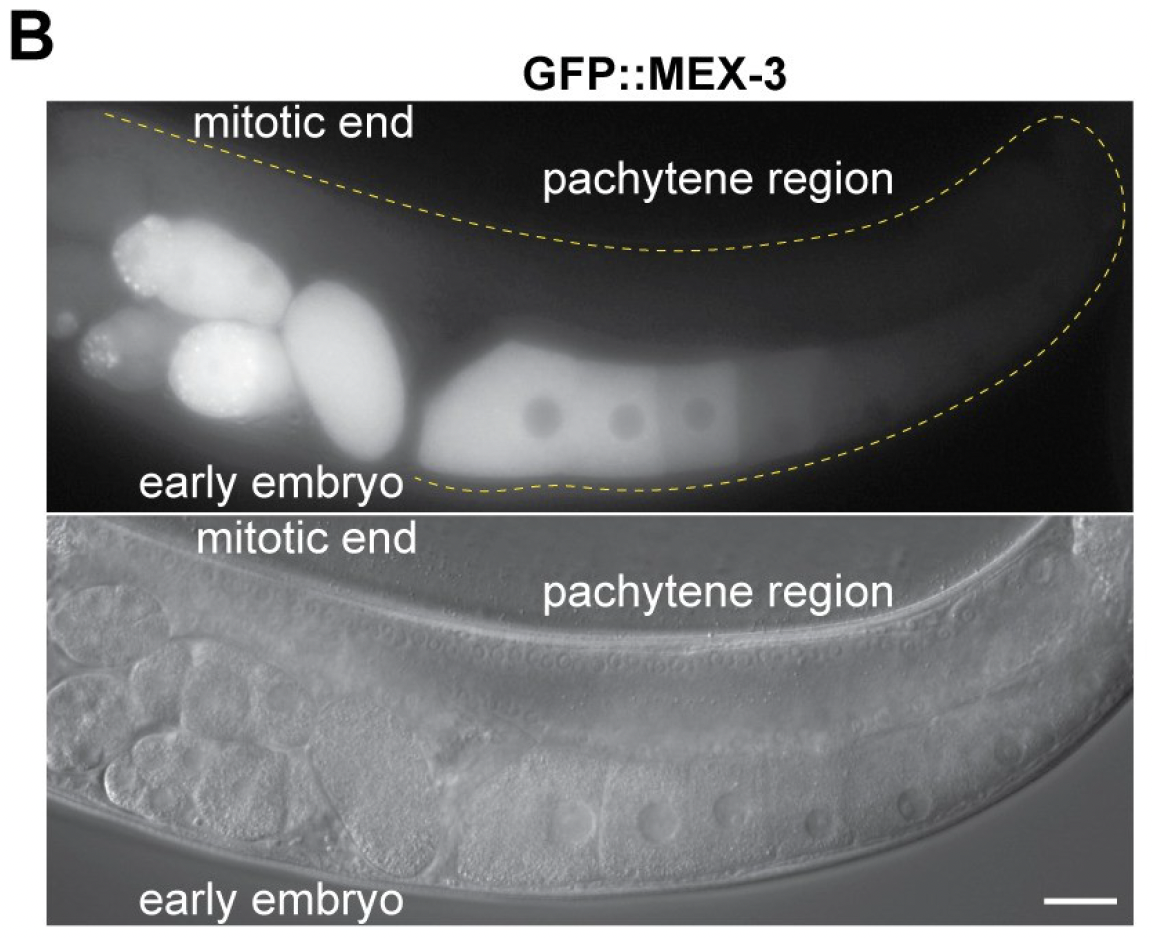
This figure from Albarqi & Ryder, 2021 shows where Mex-3 is being expressed in the C. elegans germline. Panel B shows Mex-3 protein conjugated to green fluorescent protein (GFP). The GFP signal (white) shows that Mex-3 protein is being made in oocytes and early embryos. The image beneath it is a light microscopy image of the same worm showing the germ cells without the GFP label.
Dr. Ryder and his team decided to tackle this question. They found that part of the RNA transcript of mex-3 plays an important role in regulating when and where mex-3 is expressed in the oocyte and the embryo3. The oocyte blocks the use of the mex-3 RNA before it is needed by binding it up with RNA binding proteins. These proteins prevent mex-3 from being translated into protein too early. When this sequence of the mex-3 RNA is mutated, MEX-3 protein is made too early and in the wrong places within the female germline. This aberrant usage of mex-3 RNA could potentially lead to problems with egg production, fertilization, or embryo development. Indeed, Dr. Ryder’s lab showed that ectopic production of MEX-3 protein in the female germline led to a reduction in the number of embryos produced throughout the lifespan of the worm.
Whoever fills the role of “mother” in your life, let’s take this opportunity to thank them for everything they gave us, including all the RNAs we used to get our lives started back when we were just a single cell.
To learn more about Dr. Ryder’s research, click here.