Lab Research
Retina
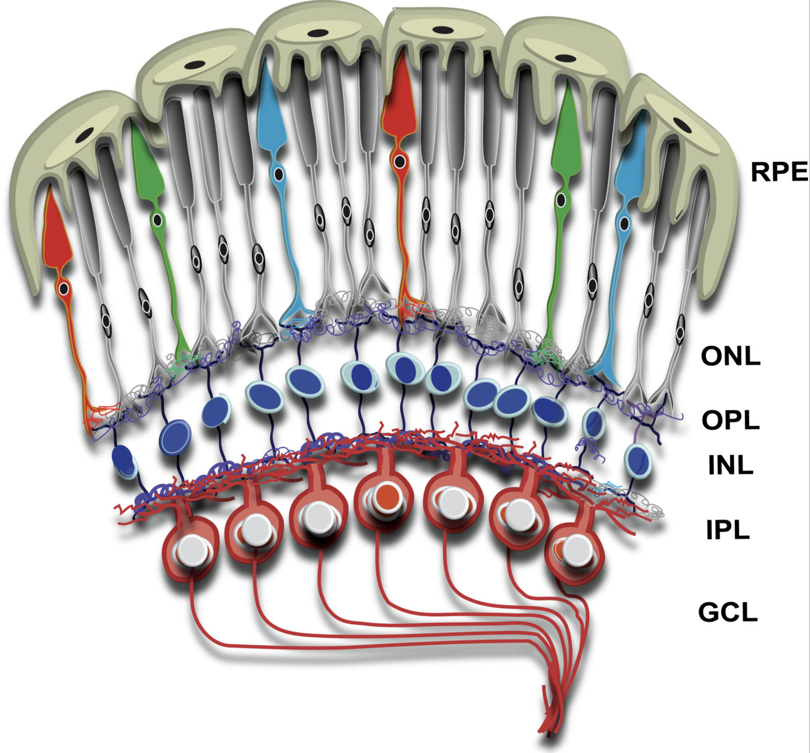
The retina is ~0.5 mm thick tissue situated in the back of the eye and is involved in the first steps of light sensation. It is a highly organized tissue consisting of six major types of neurons and one glial cell type separated by two synaptic layers, called the outer and the inner plexiform layers. Among the neurons, photoreceptors are the most abundant cell types and form the outermost layer of the retina. The tips of the photoreceptors are physically closest to the retinal pigment epithelium (RPE), which forms the outermost blood-retina barrier and is also involved in the visual cycle and periodic maintenance of the photoreceptor sensory compartment. The choroidal blood vessels overlying the RPE supply nutrients to photoreceptors.
Photoreceptors
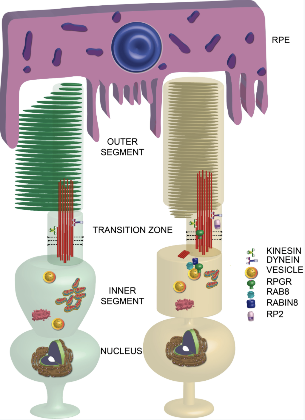
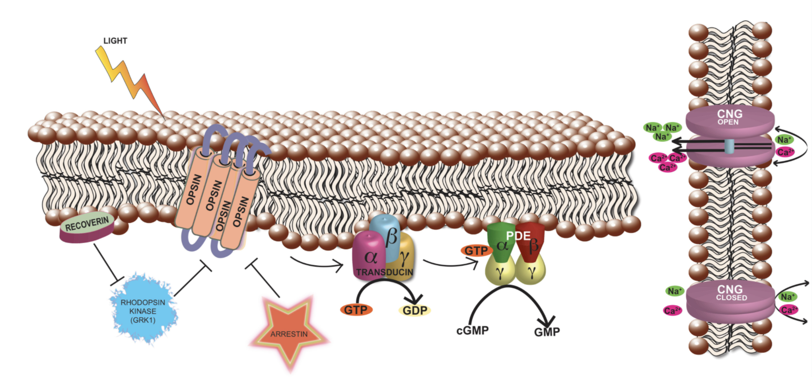
Photoreceptors are highly polarized and metabolically active neurons with a distinct compartment, called the OS, to house the phototransduction machinery. The OS is a modified sensory cilium, which contains membranous discs arranged in a coin-stack like fashion. This elegantly complex structure is devoid of any protein translation machinery; hence, the components that populate the OS are synthesized in the inner segment (IS), which contains all the necessary organelles, including the endoplasmic reticulum (ER), Golgi, and mitochondria, and transported to the distal OS. Distal to the inner segment is the cell body containing the nucleus and synaptic termini that extend into the outer plexiform layer where they synapse with the second-order neurons called bipolar cells.
For information on specific projects, click here.