UMass Ophthalmology Trainees Presenting Their ARVO Original Research
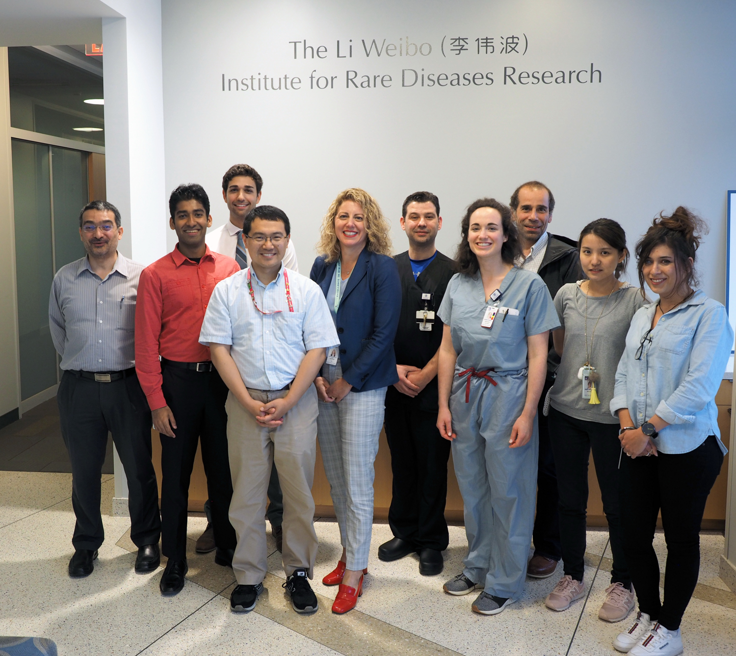
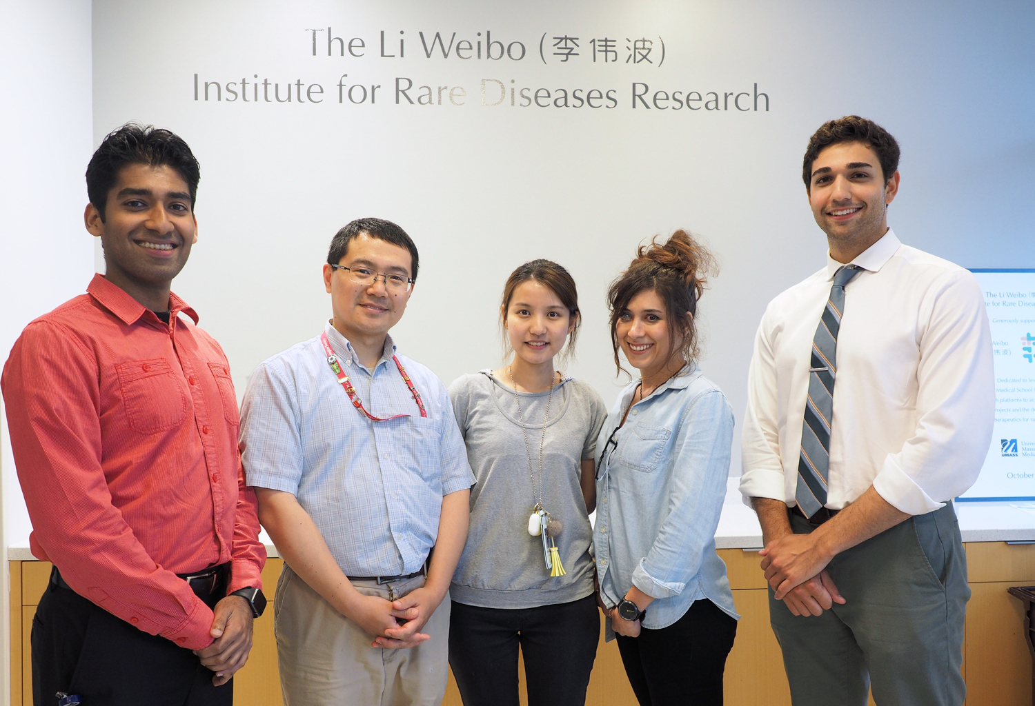
Date Posted: Sunday, June 23, 2019Several members of the Department of Ophthalmology & Visual Sciences, UMass Chan Medical School participated in the ARVO 2019 annual meeting from April 28-May 3, 2019 in Vancouver, British Columbia. The participants also presented their findings at the Ophthalmology Vision seminar series on June 20, 2019 at the UMass Chan Medical School. Here are the highlights of their presentation day:
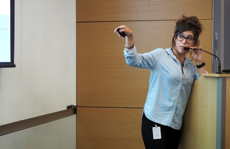
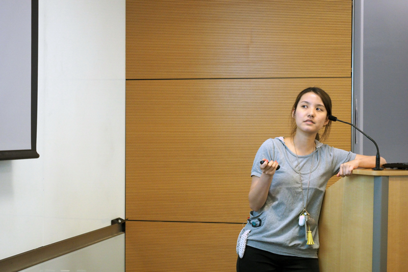
Regulation of RPGR (retinitis pigmentosa GTPase regulator) isoform expression by miRNAs: insights into retinitis pigmentosa pathogenesis (by Laura Moreno Leon).
 After obtaining my PhD in molecular biology and non-coding RNAs at the university of Nice, France, in 2017, I joined Dr. Khanna's team as a post-doctoral associate. My main goal is to understand the role of the Retinitis Pigmentosa GTPase Regulator (RPGR) isoforms in photoreceptors physiology to develop new therapeutic strategies for patients with X-linked retinitis pigmentosa (XLRP).
The project I presented at ARVO was focused on studying the precise role of the two RPGR isoforms (RPGRCONST and RPGRORF15) during disc formation and renewal in photoreceptors through a microRNAs-mediated silencing strategy of each isoform. Our results show that each isoform has a specific miRNA that regulates its expression during ciliogenesis. The two micro RNAs could thus serve as a therapeutic strategy to rebalance proper expression of the two RPGR isoforms in humans with RPGR mutations that affect only one of the two isoforms.
Isoform-specific Rpgmutant mice depict distinct roles of Rpgr isoforms in photoreceptors (by Wei Zhang)
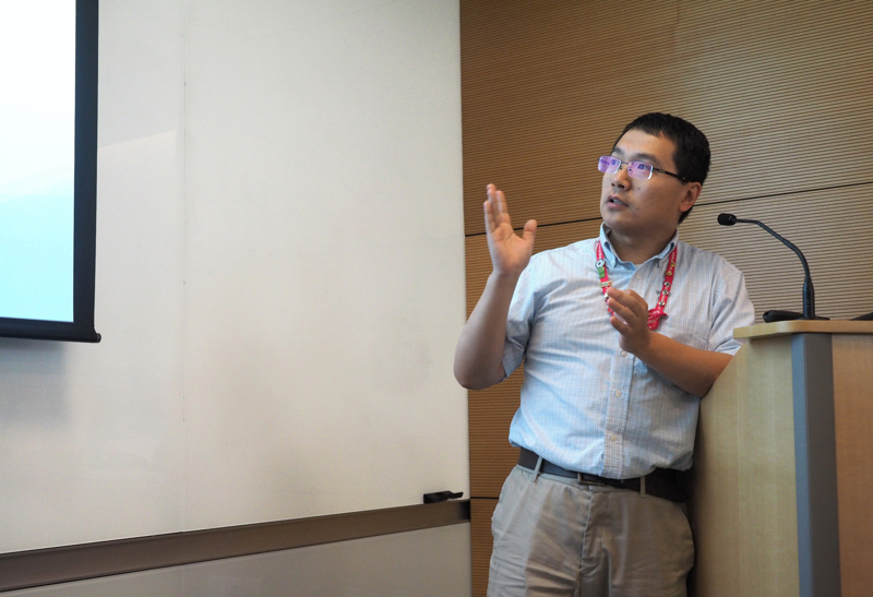 My name is Wei Zhang, I am a postdoctoral research specialist in Dr. Khanna’s Lab. My project involves the use of multiple animal models to uncover the mechanisms of CEP290 and RPGR in retinal degeneration associated mutations and design appropriate therapeutic strategies in zebrafish and mice.
The work I presented on ARVO was on understanding the precise role of RPGR isoforms in photoreceptors. To that end, we modeled a human RPGR disease-causing mutation in C57BL/6J mice by CRISPR/Cas9 strategy, which induced a premature stop codon E626X upstream of the mutational hotspot in exon ORF15 (RpgrORF15-ko). ERG analysis of RpgrORF14/15-ko mice revealed early and progressive loss of both, rod and cone function starting at ~4 weeks of age. Consistently, the mutant mice showed early cone opsin and rhodopsin mis-localization and degeneration of the photoreceptor outer segments. These studies provide new insights on the role of RPGR isoforms in regulating photoreceptor function and associated pathogenesis.
Elucidating the role of photoreceptors in AMD pathogenesis (by Shun-Yun Cheng)
 My name is Shun-Yun Cheng and I am currently a 4th year PhD student in Dr. Punzo’s laboratory. My project focuses on how photoreceptors contribute to age-related macular degeneration (AMD). We hypothesized that age-related changes in photoreceptors may lead to the disease progression of AMD. Based on this hypothesis we generated a mouse model that leads to metabolic changes in photoreceptors.
This resulted in AMD-like pathologies, including geography atrophy and choroidal neovascularization. Interestingly, the metabolic change in our mice caused also a reduction in retinal DHA levels. Dietary supplementation of DHA was able to prevent disease progression to the advanced stages of AMD, which is in line with the human studies that show a protective effect of dietary DHA on advanced AMD. We thus hope that our model will reveal new insights into the disease mechanism in order for us to develop new therapies for AMD.
Analysis of a Scribe’s Impact in an Academic Ophthalmology Clinic (by Samuel Leeman)
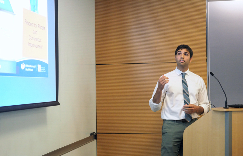 My name is Samuel Leeman, and I’ve been working for the department of ophthalmology since July 2017 as a research assistant in Dr. Shlomit Schaal’s laboratory. This coming year I will be continuing to coordinate the clinical research projects of the department as well as managing a clinical trial investigating some of the genes involved in Age-Related Macular Degeneration.
In May of 2018, I helped launch a pilot medical scribing program in the eye center as an effort to increase clinical efficiency and aid the physicians. Medical scribes have become increasingly popular in healthcare over the last decade, however, scribe impact has not been well studied outside of primary care and emergency medicine. Throughout the year we tracked metrics in satisfaction and productivity to quantify the change a scribe might bring to an academic ophthalmology clinic.
Satisfaction metrics utilized 5-point Likert scale surveys and demonstrated an increase in workplace satisfaction among the four participating physicians, with no significant change patient satisfaction due to a high baseline from all respondents. Productivity metrics analyzed physician RVUs and visit length times, showing a small increase in the average RVU per session with a scribe, however, future studies will need to change the physician’s schedule based on when they are with a scribe to observe more accurate RVU changes.
Analysis of Vascular Changes in Cerebrovascular Accident Patients via OCT Angiography (by Sahil Shah)
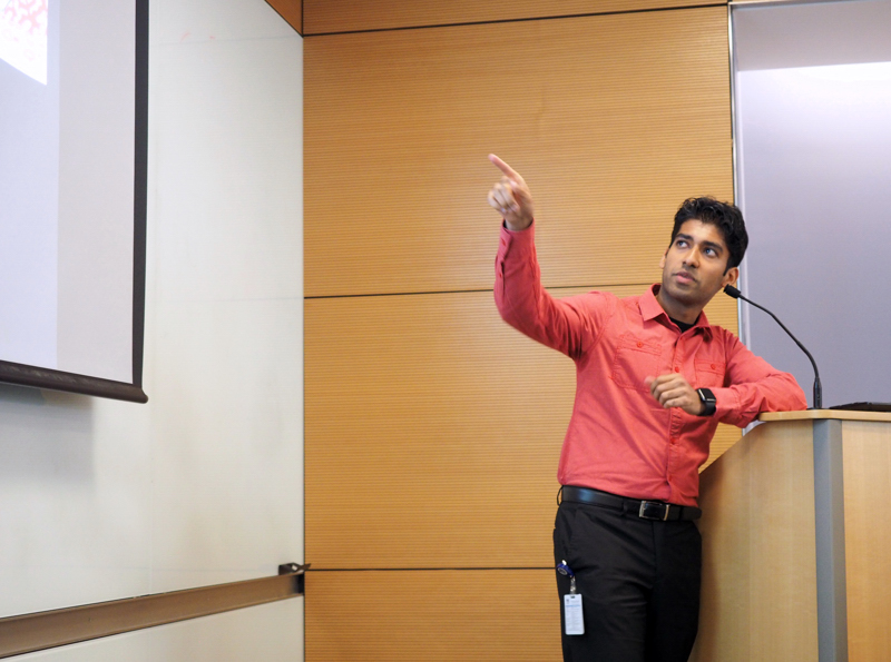 My name is Sahil Shah and I am a rising 2nd year medical student at UMass Chan Medical School. Born and raised in Winchester, MA, I’ve stayed local my whole life, attending Tufts University for undergrad and working as a medical scribe in my gap year before starting medical school. Ophthalmology has been a particularly interesting field for me to explore, and I’m excited to share the work I have done so far!
My project, titled Analysis of Vascular Changes in Stroke Patients via OCT Angiography, aims to determine whether there are any detectable changes in retinal blood vessels in patients who have had a stroke, and whether we can use imaging of the eye as a diagnostic tool to predict the likelihood of having a stroke before it occurs.
Our analysis of OCT angiography images across 3 layers of retinal vasculature (superficial, deep, and choriocapillaris) reveals significant changes in density (vessel architecture) and intensity (blood flow) of vessels on imaging. Interestingly, the choriocapillaris layer had the most significant decreases in vessel intensity and an increase in vessel density. Through further data collection and new analysis methods we hope to gain further insight into what these vascular changes could mean for diagnosis of stroke on eye imaging.