The New OCT Angiography machine brings excitement to the Ophthalmic Clinicians and Researchers
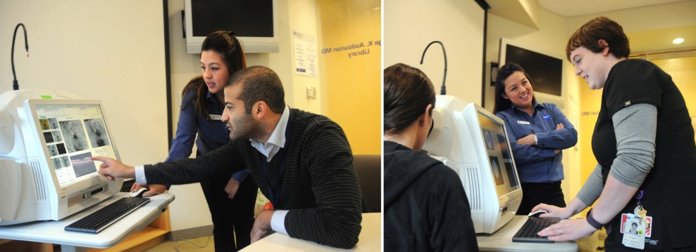
Date Posted: Monday, October 31, 2016A new technology advancement OCT machine (Optical Coherence Tomography) finally becomes part of the Department of Ophthalmology & Visual Sciences at the University of Massachusetts. In contrast to the previous versions of OCT, this machine allows clinicians to observe fine details of the retinal vasculature both normal and abnormal. This advanced, non-ivasive OCT machine with no dye required already provides a huge advantage for patients by avoiding discomfort and tedious procedures of dye injection.
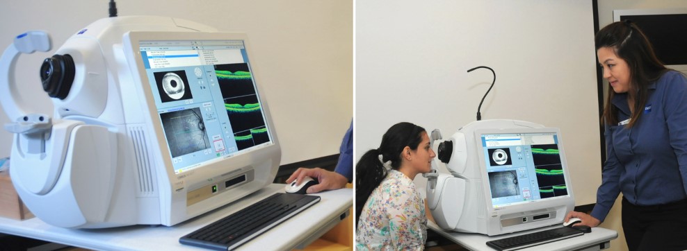
The New OCT machine uses a unique algorithm that detects movement of red blood cells as a contrast. The algorithm can also separate and visualize different layers of the retina and choroid vasculature. The software has a color coded option, that allows depth visualization of retinal blood flow. So what does that mean to clinicians? This exciting new technology gives us the opportunity to look at various retinal diseases in a completely different perspective, creating novel research opportunities for scientists to understand further the origin and mechanism of pathologies.
OCT Angiography can be of great benefit to common diseases such as areas of choroidal neovascularization in AMD, micro-aneurysm and ischemia in diabetic retinopathy, occlusion and abnormalities central and branch retinal vein occlusions, and abnormalities of optic nerve vasculature. Clinician Scientists in the UMass Ophthalmology Department are looking forward to make new discoveries that will change the way we observe common diseases and be able to better treat patients.