

Photo: Bryan Goodchild
Using computational methods he developed, first year MD/PhD student Connor J. Lewis found that results of a Phase I/II clinical trial for a single infusion of an adeno-associated virus (AAV) gene vector for Type II GM1 gangliosidosis showed improved neuroimaging patterns following treatment. The results of the study, co-authored by Lewis and published in the New England Journal of Medicine, also showed improved biomarkers and stable or improved rates of developmental deterioration in some patients.
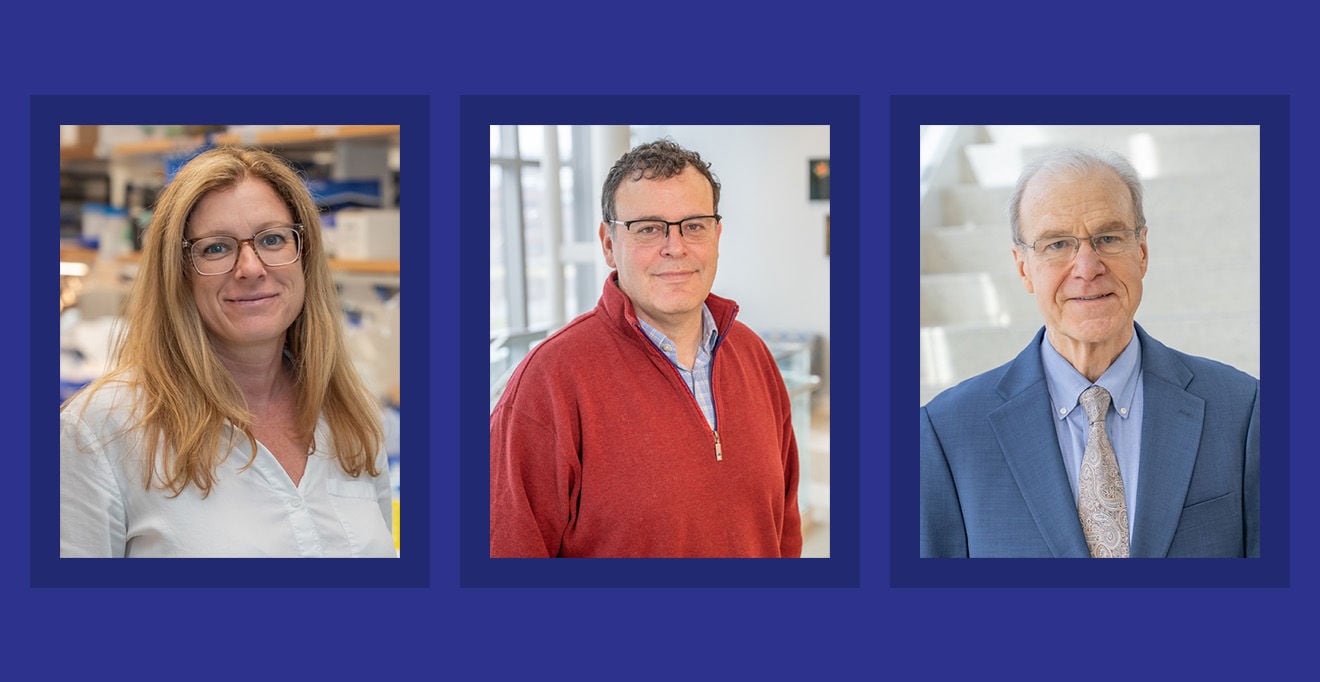
Led by Cynthia Tifft, MD, PhD, deputy clinical director at the National Human Genome Research Institute (NHGRI) in Bethesda, Maryland, the trial used an AAV vector developed by Miguel Sena-Esteves, PhD, associate professor of genetic & cellular medicine, and Heather L. Gray-Edwards, DVM, PhD, associate professor of genetic & cellular medicine, at UMass Chan Medical School.
Lewis began working with Dr. Tifft at the NHGRI as a post-baccalaureate intramural research trainee after graduating with a bachelor’s degree in biomedical engineering from Virginia Commonwealth University in 2023. His work at the NHGRI included collaborating with Dr. Sena-Esteves and Dr. Gray-Edwards, as well as Terence R. Flotte, MD, the Elisabeth Chair for the Dean of Medicine, provost and executive deputy chancellor, and Allison Keeler, PhD’14, assistant professor of genetics & cellular medicine, at UMass Chan, on several gene therapy studies for GM1 and GM2 gangliosidosis.
“My contribution to the study was in advanced neuroimaging analysis,” said Lewis. “I developed computational methods of interpreting diffusion tensor imaging, which allow for the viewing of changes in neuronal pathways. In this study, we were able to use these techniques to map neuronal pathways in patients before and after treatment and compare them for neuronal loss or growth.”
Diffusion tensor imaging is an advanced MRI technique that compares and combines multiple images to map exact white matter segments showing neuronal injury or change. It improves upon conventional imaging by specifically tracking the directional movement of water molecules in the brain through diffusion. By following changes to how water moves in the brain, scientists and clinicians can generate quantitative data tracking disease progression in patients with multiple sclerosis, ALS and other conditions such as GM1 and GM2.
Typically, neuroimaging has shown consistent declines in neuronal fiber-tract number and volume in patients with GM1 gangliosidosis. Patients treated in the study, however, showed gains in neuronal fiber tracts up to three years post-treatment, suggesting a move in a positive direction for treatment.
“What Connor has done is incredibly important for the field. This technology shows the impact gene therapy can have at the most fundamental level,” said Sena-Esteves. “It gives a representation of an effect we often struggle to see.”
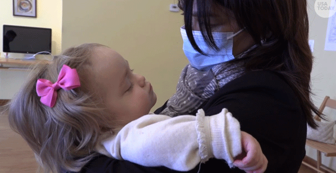
GM1 gangliosidosis is a rare and fatal lysosomal storage disorder caused by a mutation in the GLB1 gene that leads to a deficiency in the beta-galactosidase enzyme. This deficiency causes toxic accumulation of gangliosides in neuronal cells, leading to progressive neurodegeneration, developmental regression and death. Patients diagnosed with Type II GM1 gangliosidosis have an average lifespan of 20 to 40 years.
It was Lewis’s collaboration with Sena-Esteves, Gray-Edwards and Dr. Flotte that led him to apply to UMass Chan’s MD/PhD program.
“I had strong mentors in place at UMass Chan. For students interested in gene therapy, there are exciting things happening and UMass Chan is the place to be,” said Lewis. “I knew that coming here I’d be able to continue on this path of researching gene therapy, neuroimaging and rare diseases.”