Umbilical cord development and physiology
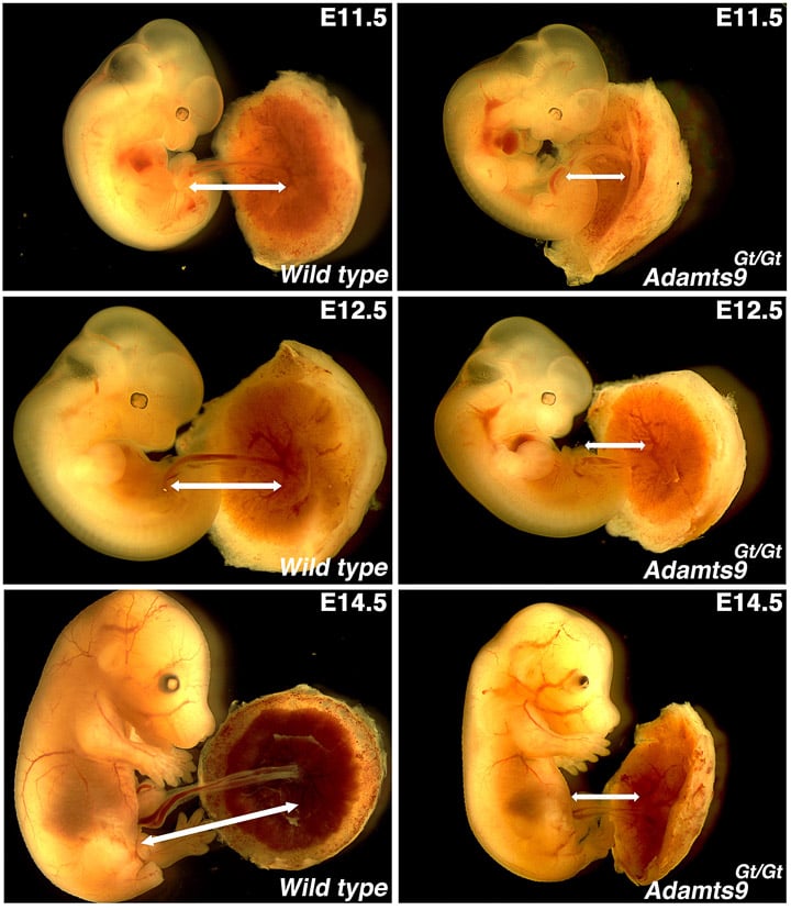 The umbilical cord is a fascinating extraembryonic vascular organ which is crucial for embryonic survival. Yet, its development is mostly ignored and very little is known about the cellular and molecular pathways governing cord development and its physiology. The umbilical blood vessels (typically containing two arteries and one vein), are engulfed by a protective connective tissue known as the Wharton’s jelly, preventing the accidental kinking of the vessels and cessation of circulation leading to sudden fetal death. As the embryo rapidly grows, this protective connective tissue and the ECM surrounding the umbilical vessels must be continuously turned-over for growth and expansion of the cord vessels. In previous work, we identified ADAMTS9 as one of the first genes to be characterized crucial for umbilical cord development (Nandadasa et al., Cell Reports 2015). We discovered that mouse umbilical cord smooth muscle cells (SMCs) undergo a crucial reorientation process at E12.5 which is critical for rapid umbilical cord elongation. Adamts9 mutant SMCs fail to undergo reorientation and elongation of the umbilical cord is abrogated at E11.5 (Image-1, Nandadasa et al., Cell Reports 2015) resulting in fetal death at E14.5.
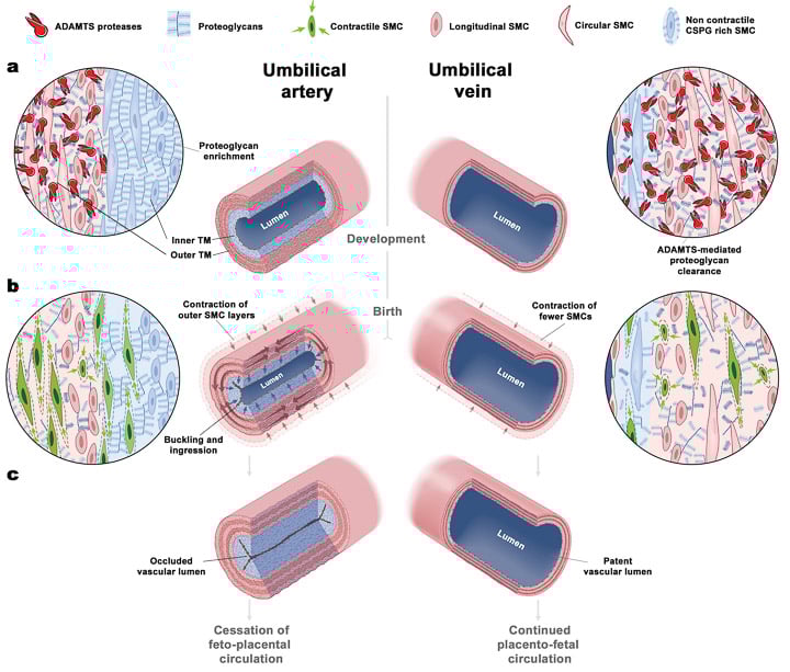
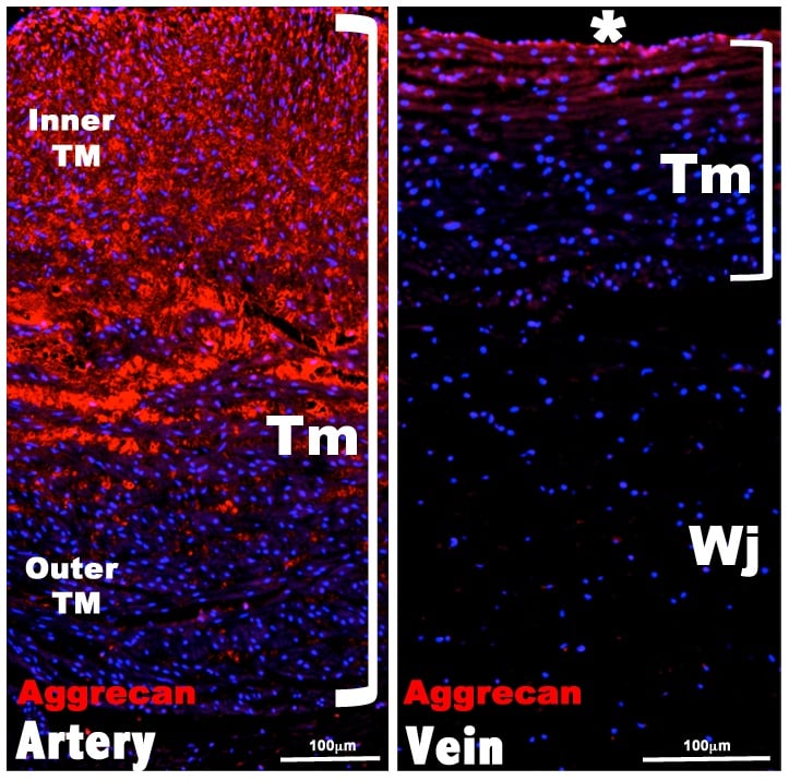 By the end of gestation, umbilical cord vessels develop an amazing physiological mechanism for ensuring rapid vascular occlusion preventing exsanguination of the newborn when the cord is severed at birth. The exact cellular and molecular mechanism underlying this process was not previously known. In a groundbreaking study, we revealed that aggrecan, a chondroitin sulfate proteoglycan known to be unique to cartilage, is crucial for this process (image-2, Nandadasa et al., eLife 2020). We discovered that ADAMTS proteases expressed by the vein and outer tunica media of the arteries ensures accumulation of aggrecan is limited to the inner tunica media of the artery. At birth, the outer tunica media undergoes rapid constriction resulting in the luminal buckling and collapse of the proteoglycan rich inner SMC layer, instantly occluding the vascular lumen of the arteries (image-3). The umbilical vein does not have this specialized proteoglycan rich inner layer and does not undergo rapid occlusion, allowing for placental infusion. We showed that this mechanism is used by many mammals other than humans and mice (such as elephants, walrus, rhinoceros, chimpanzees, bonobo etc.), and also theoretical proved our model by mathematical modeling in collaboration with the Humphrey lab at Yale University.
In future studies, we will continue to study the development and physiology of this important organ and characterize the role of primary cilia and hedgehog signaling during umbilical cord morphogenesis, elongation and occlusion.