


The Morrison laboratory, part of the Department of Microbiology, is located in the Sherman Center of the UMass Chan Medical School in Worcester, MA.
The Morrison laboratory is interested in basic mechanisms of infection by enveloped RNA viruses. We have focused on the entry of viruses into cells as well as progeny virus assembly and release from infected cells. These studies have led us to also explore the role of virus glycoprotein conformation in stimulating optimal protective immune responses in animals important for vaccine development. Our focus has been on Newcastle disease virus, a paramyxovirus, and respiratory syncytial virus (RSV), a metapneumovirus, both of which are negative stranded, non-segmented, enveloped RNA viruses. More recently, we have initiated studies of the coronavirus SARS-CoV-2, a positive stranded, enveloped RNA virus. Our studies of virus assembly have led us to characterize the release of virus-like particles (VLPs) from cells expressing only the major structural proteins of Newcastle disease virus. These VLPs provide an excellent system to explore requirements for paramyxovirus assembly. They are also excellent vehicles for expression of a number of different viral glycoproteins and different conformational forms of viral glycoproteins in a virus-sized particle. These particles can be used as vaccines to explore immune responses stimulated by different glycoproteins in experimental animals. Our current projects focus on the conformation of the viral fusion proteins in stimulating optimal titers of protective neutralizing antibodies in experimental animals (mice, cotton rats, and primates) with the goal of optimizing design of human vaccines.
For additional information, please contact Trudy Morrison (trudy.morrison@umassmed.edu).
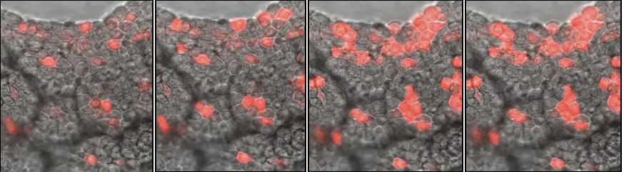
Spread of RSV infection in mouse lungs: Recombinant RSV with a RFP gene inserted into the genome was used to infect mouse lungs. Using two photon microscopy, photos were taken of lung slices at different times after the beginning of infection. The increase in red fluorescent cells indicates the spread of the virus infection through the tissue. (M. Sanderson and T. Morrison)