Click here to access Microscopy Calibration Survey
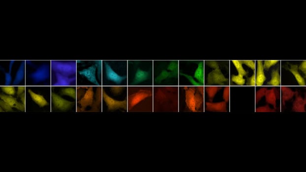
Our biological research interests lay in the function of the cell nucleus, in particular focused on elucidating mRNA trafficking in the nucleus, its transport across the nuclear membrane and the interplay of different transport pathways. Our approach aims at understanding the basic mechanisms of nuclear function especially as several diseases have been directly linked to defects in mRNA nucleocytoplasmic transport. Our ambition is to reproduce the biochemical and physical reactions elucidated in the test tube directly in the cell. To do so we use genetic labels for proteins and RNA combined with viral gene delivery and genome editing tools (CRISPR) to make these, often weak and highly transient reactions visible using fluorescence microscopy. In particular we actively develop Single-molecule live-cell real-time (SMRT) quantitative microscopy.
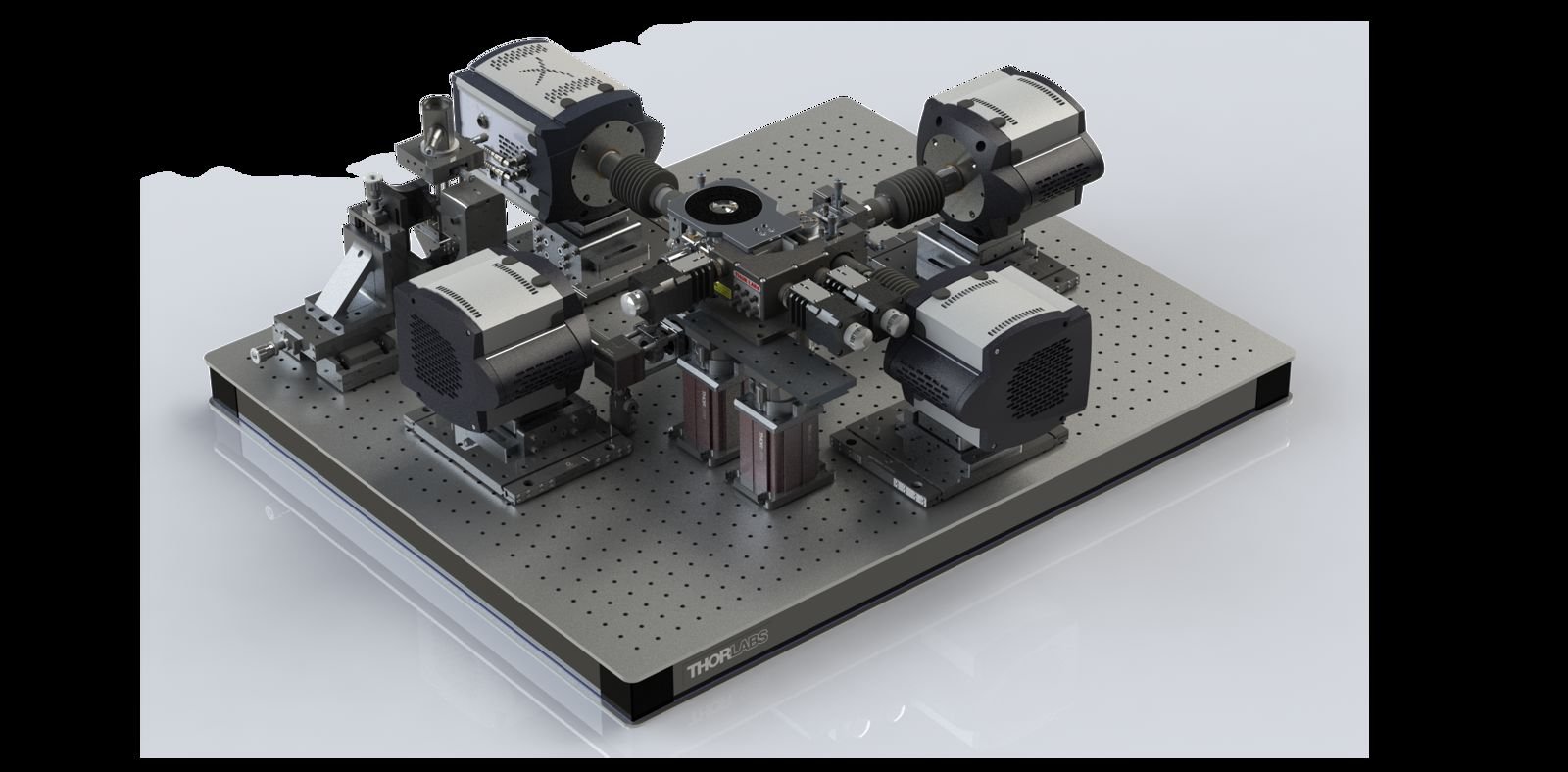
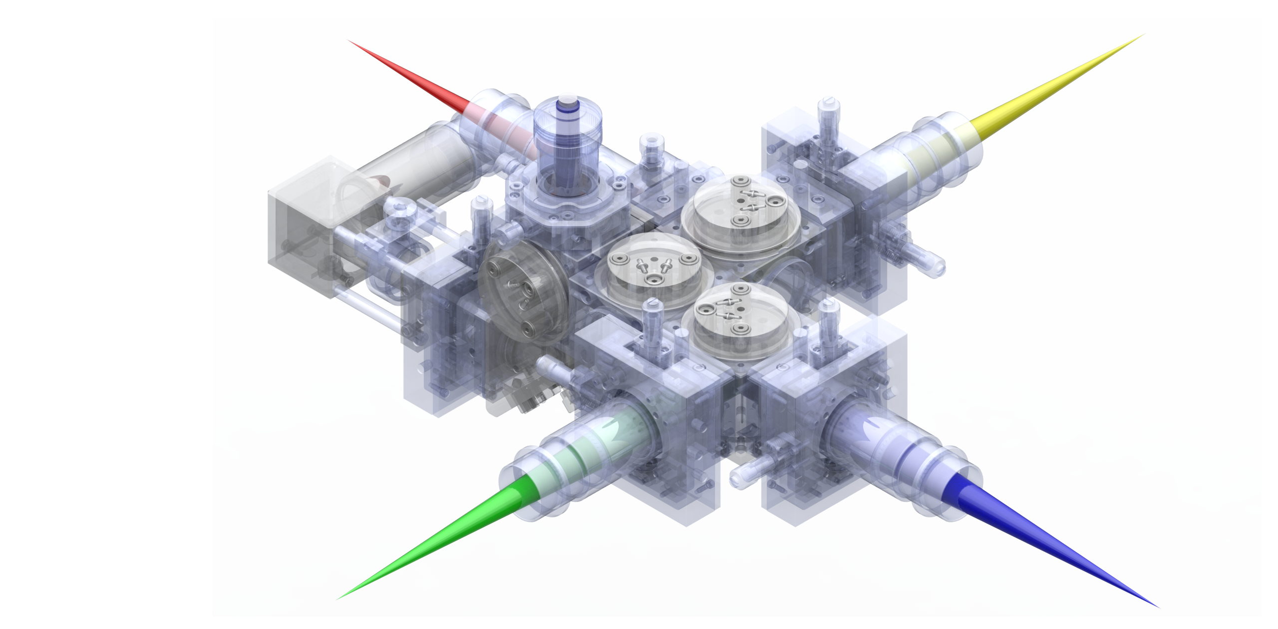
With SMRT microscopy it is possible to visualize what, where, when and how fast a process is happening inside the cell. SMRT microscopy can reproducibly detect weak fluorescent signals within a cell with sub-diffraction limited precision at high temporal acquisition frequencies. The images provided by SMRT microscopy contain ample information on the reaction kinetics, temporal fluctuations, efficiencies and spatial location of molecular interactions that can be quantitatively measured. We do build our own microscopes and work on SMRT microscopy in 3D and spectrally multiplexed. SMRT microscopy demands advanced prototype designs, but beside optics retrieving information correctly from the images we record is a major challenge. However, in the grand picture all the technology and tools we develop have only one aim: to do what matters the most to us - exploring the stochastical molecular landscape of the living cell.