Services
The Laboratory of Molecular Imaging probes has about 20 years of experience in the field of in vivo molecular imaging.
We perform fee-for-service development of small molecules and nanoparticles designed for optical, photoacoustic and MR imaging. The latter include both paramagnetic and superparamagnetic imaging probes. We are also equipped to perform purification, formulation and initial testing in vitro and in vivo.
We previously performed the following services for the several academic and industry clients:
- Synthesis, purification, testing and formulation of small-molecule peroxidase-imaging probes (Universities in Germany, Korea and Israel)
- Synthesis of near-infrared fluorescent polymer-based probes (Company in Seattle WA area)
- Synthesis of superparamagnetic antibody-linked nanoparticles (Company in Washington DC area)
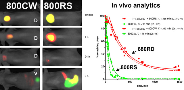
- Imaging of dual-labeled near-infrared fluorescent peptides and polymers in mice after local injection (Company in Seattle WA area).
- Formulation of poorly soluble drugs for systemic administration (Company in Bothell WA area)
We are always ready to discuss your custom synthesis R&D project to determine the best options you have for developing a custom imaging probe and sensor.
 |
| Left: dorsal (D) and ventral (V) images of mice injected with a complex between a macromolecular drug carrier (hydrophobic core HC-PGC labeled with IRDye 680) and either IRDye 800CW (more hydrophilic) or IRDye 800RS (less hydrophilic). Near-infrared images were collected in two channels (red = IRDye680 dye (700 nm fluorescence channel); green= IRDye 800 (800 nm fluorescence channel) at time intervals indicated on the right. Monoexponential curve fitting showing fluorophore elimination from the SQ injection side. Red traces – HC-PGC-680 elimination kinetics, green traces – 800 nm fluorophore elimination kinetics. Fitting results are shown in the legend. Copyright: European Society for Molecular Imaging (ESMI, 2018). |