


Fluorescent Imaging of Cellular Fluxes
Seeing is Believing!The rapid bursts of ions exiting cells are vital for many physiological processes. The challenge is visualizing these multiple events from cells where the coordinated release of ions is the basis of neuronal, cardiac, and muscle excitability and where perturbation results in human disease. Conventional electrical methods either report on total ion flow from a particular cell (electrophysiology) or an ion concentration at a specific location (ion-sensitive electrodes); however, neither approach provides the spatiotemporal resolution to globally image and measure efflux from the entire landscape of a cell, an intact circuit, tissue, or brain. Therefore, we have been developing a novel cell surface approach to fluorescently visualize ions and metabolites exiting cells. |
|

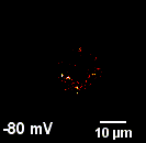
Proton depletion @ -120 mV |
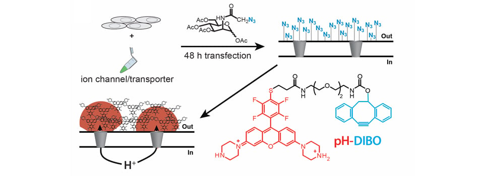
Real-time Imaging of Cellular Proton FluxesWe are using glycocalyx engineering (pioneered by Bertozzi and co-workers) to covalently label the cell surface with small molecule fluorescent pH sensors (cartoon). Using voltage-clamp fluorimetry to simultaneously control and visualize proton transport, we observe robust fluorescent signals that correspond to cellular proton fluxes in both directions—outward and inward. The real-time kinetics of proton accumulation and depletion at the cell surface directly serves as a readout of ion channel and membrane transporter activity. Using our approach, we have observed proton transport through voltage-gated proton channels, chloride/proton antiporters, and omega proton fluxes associated with human disease. |
Proton accumulation @ 100 mV |
What will we see next?
Hopefully everything! We can image four different fluorphores at 400 images per second on our voltage-clamp fluorometry set up thanks to a CoolLeD pE-4000, quad filter sets from Chroma, and a sCMOS Zyla camera from Andor. We are now imaging proton transport through non-electrogenic transporters that cannot be detected using electrophysiology. In addition, we are branching out beyond protons: potassium, lactate, and glucose are all on our radar. Moreover, we are developing a new technology to visualize extracellular ion concentrations for any ion or metabolite. The ultimate goal is to enable visualization of extracellular ion and metabolite activity in tissues and living animals.