

Instrumentation
 |
 |
 |
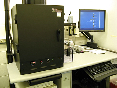
IVIS-100 (Perkin-Elmer) |
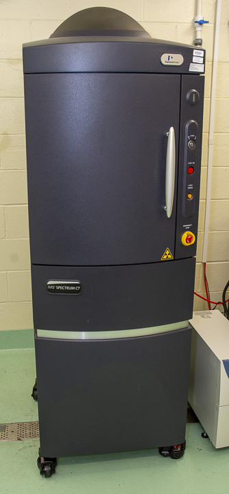
IVIS Spectrum CT (Perkin-Elmer) |
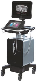
Vevo 3100 Ultrasound (FujiFilm VisualSonics) |
Both IVIS cameras detect bioluminescence and fluorescence contained within solutions, petri dishes, culture plates, cells or live animals.
IVIS-100 - Animal Facility, A-level, Room SA-443
The IVIS-100 has 4 excitation/emission fluorescent filters and covers the excitation range of 445-750 nm and emission of 500-875 nm. The chamber holds 1-5 mice or 1-3 rats in a single view. Acquisition time is about 1-3 minutes; therefore, useful for high-throughput screening. The system provides 2D imaging with photo overlay and tools for quantification of the surface radiance of the source with Living Image Software.
IVIS Spectrum CT - Animal Facility, A-level, Room SA-414
The IVIS Spectrum CT has 10 excitation and 18 emission fluorescent filters, along with spectral unmixing to facilitate the use of multiple signals within the same subject. The system provides both 2D and 3D imaging (with CT). The 3D aspect calculates the X, Y, Z location and brightness of the source in the subject at depth and quantifies a depth corrected signal intensity. CT registration provides anatomical registration.
Each camera is equipped with a gaseous vaporizer for anesthesia. Oxygen and isoflurane are provided.
Vevo 3100 High Resolution Ultrasound System (VisualSonics) for small animal models - Animal Facility, A-level, Room SA-414
The instrument is available to investigators through the Optical Core. It can be used for general organ imaging and cancer models.
Applications include:
- Image guided needle injection
- perfusion tracking
- reproductive health evaluation
- organ 3D volume
- tumor verification and sizing
To learn how to get started and to arrange training contact Alice Truong or Mary Rusckowski.
Use On-line Calendar to reserve time.
INSTRUMENT USE
- Optical imaging of live animals requires IACUC approval in an active animal use protocol. Please check that optical imaging and related procedures are included in your animal protocol.
- Equipment can be used without assistance after training by Core staff.
- Core staff is available on request to assist with operation of the camera for your studies. For assistance please contact Yuzhen Wang, Core Manager.
- To reserve a block of time, use the on-line calendar, link is below, “Make a Reservation”. Be sure to sign-up in advance. Cancellation of reserved time is through the same site.
- An in-room log book is also used to enter your arrival and departure times.
Certified users that have been trained can schedule time using this Make a Reservation link.
For assistance navigating the on-line calendar, to schedule assisted time or training contact:
Alice.Truong@umassmed.edu
508-856-3791.