
 |
|
|
Andrei Korostelev, PhD |
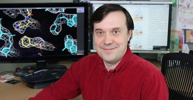
Cryo-electron microscopy is allowing scientists to answer questions about cellular processes that are essential to life, as demonstrated by a study led by UMass Medical School structural biologist Andrei Korostelev, PhD. Published in Nature, the study answered basic questions about the mechanism by which ribosomes, the molecular machines responsible for reading genetic information, enable the synthesis of life-sustaining proteins in every cell.
“The transfer RNAs and ribosome decoding centers that read their genetic instructions are almost the same for all life on earth—that’s why we are very excited by this discovery, which explains the accuracy of genetic code reading,” said Dr. Korostelev, associate professor of RNA therapeutics and biochemistry & molecular pharmacology. “Cryo-EM was absolutely critical for us to distinguish between the ribosome’s reading of correct and incorrect tRNA.”
One of the original proponents for bringing the Massachusetts Cryo-Electron Microscopy Facility to UMMS, Korostelev studies how cells utilize ribosomes to make the proteins that are their building blocks. Two cryo-EM systems comprise the newly built research core, which opened last fall at UMMS. It is New England’s first installation of the transformative technology.
Cryo-EM is a breakthrough that allows researchers to visualize the detailed structure of cells, viruses and proteins at near-atomic resolution. In the experiments detailed in Nature, investigators visualized with cryo-EM the distinct steps in which the ribosome’s parts move and align to ensure the correct decoding that is essential for normal cell growth and development. Ribosomes, transfer RNAs, messenger RNA codons, nucleotides and amino acids each have distinct roles that were clearly seen with cryo-EM.
Understanding these fundamental molecular mechanisms may ultimately have implications for drug design. For example, many antibiotics that kill bacteria do so by forcing miscoding in bacterial ribosomes. New antibiotics could be designed to disrupt coding accuracy in bacteria that have become drug resistant.
The Korostelev lab co-authored the paper with Nikolaus Grigorieff, PhD, a cryo-EM pioneer with whom Korostelev has been collaborating since 2010. Dr. Grigorieff, group leader at the Howard Hughes Medical Institute Janelia Research Campus, was the keynote speaker at the opening of the cryo-EM facility UMMS last fall.
“The state-of-the-art technology that enabled this research is now available at UMass Medical School and we will continue our studies here,” said Korostelev.
The research team is now developing time-resolved cryo-EM, an even more advanced technique than that used in the current study in which more steps of a process can be captured in the proper order to further understand translation.
Related stories on UMassMedNow:
Cutting edge UMMS Cryo-Electron Microscopy facility opens
Massachusetts Facility for High-Resolution Cryo-Electron Microscopy opening at UMMS
Massachusetts Life Sciences Center announces $5M award to UMMS for Cryo-Electron Microscope