

Immuno-Electron Microscopy
|
|
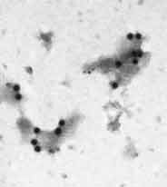
Double labeled cell membrane fragments. large gold beads- glucose transporter, Glut4; small gold beads, vimentin. (Courtesy of Drs Michael Czech and Adilson Guilherme, Biochemistry Molecular Biology, UMass Chan Medical School) TEM |

Immuno-electron microscopy is used to localize molecules at the ultrastructural level by labeling them with specific antibodies. The antibodies are visualized by electron-opaque markers (colloidal gold particles) attached to them. The effect is to produce an electron-dense label at the site of the antigen-antibody reaction. When the antigen is located inside of the cell, then transmission electron microscopy is required to see it. The labeling can be done pre-embedding or post-embedding (on thin sections).
When the antigen in question is located on the surface of the specimen, scanning electron microscopy can be used to see it. The electron dense label can then be viewed using the back scatter image on an appropriately equipped microscope.