

Routine Scanning Electron Microscopy
 |
 |
 |
") |
| False colorized scanning electron micrograph of two macrophages (purple)phagocytizing Glucan Encapsulated RnaiParticles (GeRP, green). Courtesy of Czech's Lab, UMass Chan Medical School. |
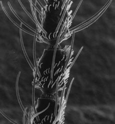
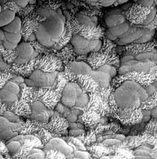
Scanning electron micrograph of an antenna of female black fly. Courtesy of Dr Robert O'Connell, Physiology, UMass Chan Medical School. | Scanning electron micrograph of a mouse trachea. Courtesy of Dr Greg Pazour, Cell Biology, UMass Chan Medical School. | Scanning electron micrograph of an ant knee taken with ESEM. Dr Greg Hendricks, Cell Biology, UMass Chan Medical School. |
Scanning electron microscopes (SEM) offer superior performance compared to light microscopes, particularly in resolution (single nanometer resolution in High vacuum mode), depth of field (up to 500 time that of a light microscope), and microanalysis (Electron Dispersive x-ray analysis and Wave Length Dispersive X-ray analysis).
A SEM can form an image using a variety of signals:
- secondary electrons emitted from the surface of the sample offer the best resolution and contain topographical information.
- X-rays produce the best information about the sample composition but have poor spatial resolution.
- backscatter electrons (atomic contrast) offer information about the atomic densities of the sample, provide medium resolution (down to several nanometers) but carry significant but non-specific compositional information.
- By simultaneously mixing these signals the investigator can produce images that contain both compositional and topographical information with astonishingly high resolution.
However, modern variable pressure scanning electron microscopes (microscopes that can operate at Low vacuum or even environmental pressures) can finally answer the question, “What does it look like in its natural state?” It was exactly this question that lead to the development of the Environmental Scanning Electron Microscope (ESEM). To do this there were two serious technical issues to solve. First, the vacuum environment of the gun and column had to be separated from the environment of the specimen chamber. And second, a new and very robust secondary electron detector that could function in a non-vacuum environment had to be developed. The solutions to these problems lead to the development of the modern “state of the art” ESEM.
In all modes of operation, the primary electron beam generates secondary electron emission at the sample surface but instead of collecting and electronically amplifying is signal in the detector, now in the ESEM mode environmental secondary electrons are generated in the water vapor surrounding the sample itself. Magnification is created in the same way in all modes of operation. The size of the raster scanned on the surface of the specimen is much smaller than the surface of the viewing monitor. Therefore, the final picture is a magnified image of the surface of the specimen.