

Room Temperature TEM
 |
 |
|
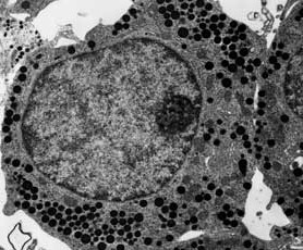
Thin sectioned epoxy embedded cell
from the pituitary gland.
|
Thin section of retina in mouse eye showing a cross section of cilia in photoreceptor. |
Conventional room temperature transmission electron microscopy requires the fixation and sectioning of cells and tissues. Tissues, organelles, cell cultures, etc. are fixed with aldehydes and osmium tetroxide, dehydrated through a graded series of alcohol to remove water, and finally embedded in a plastic resin. The resin-embedded material is sectioned using either glass or diamond knives. Sections can be either cut thick (0.5 to several micrometers) for light microscopy or ultrathin (60 to 90 nm) for conventional transmission electron microscopy.
If the structure of interest is required to be in a particular plane, or the resercher is interested in a specific cell type in a tissue then thick sections of the material are used to orient or to find the area of interest under light microscopy, and then the blocks are re-trimmed and thin sectioned.
Correlative light and electron microscopy can also be done, but the cells of interest are tracked manually at the light level, and processed through to TEM. This technique is informative but time consuming.