

Negative Staining
|
|
|
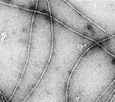
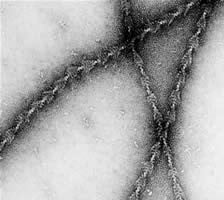
Negative staining is a simple and rapid method for studying the morphology and ultrastructure of small particulate specimens. Samples stained in this manner show great structural integrity because the stains used not only delineate the ultrastructure, but also act as a fixative, thus protecting samples from irradiation damage by the electron beam. Negative staining also reduces the surface-tension forces at the air-liquid interface, thus reducing shrinkage and specimen collapse. These effects are promoted by the heavy metal salts used in negative stains. While negative staining is one of the oldest techniques for studying the ultrastructure of particulate samples at the electron microscopy level, it has never been surpassed by any other technique for determining the surface structures, size, and shapes of specimens such as viruses, bacteria, and macromolecules, with resolution down to the 2-nm level.