
 |
|
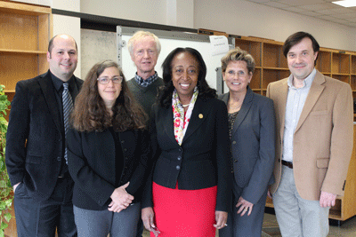
| From left, UMass Medical School’s Brian Kelch, PhD; Celia Schiffer, PhD; Roger Craig, PhD; Jean King, PhD; Susanna Perkins, and Andrei Korostelev, PhD, attend the announcement of a $5 million grant from the Massachusetts Life Sciences Center for a high resolution cryo-electron microscope at UMMS. |
The Massachusetts Life Sciences Center announced a $5 million grant to UMass Medical School for the purchase of a high resolution cryo-electron microscope (cryo-EM), bringing this breakthrough imaging technology to a campus poised to usher in the next generation of drug design and discovery. UMMS and Harvard Medical School have jointly committed operating funds to support the new Titan Krios microscope, which will be the centerpiece of the new cryo-EM research core, located on the UMMS main campus in Worcester. An innovative operating plan will encourage use of the microscope by researchers at other universities and in the biotech community, bringing this important new tool to laboratories and biotech firms across the region.
High resolution cryo-EM is a critical new technology for studying the relationships between structure and function in large molecular complexes within cells. Recent technological advances make it possible to see molecules and structures with unprecedented clarity, opening up vast new opportunities for the study of the key biological structures important to understanding disease, genetic disorders and bacterial and viral pathogenesis. Cryo-EM allows the observation of specimens that have not been stained or fixed in any way, showing them in their native environment, in contrast to X-ray crystallography, which requires crystallizing the specimen.
One of the most important new tools for drug design and development, cryo-EM will play a critical role in identifying likely therapeutic approaches for a broad range of diseases, particularly neurological disorders such as Alzheimer’s, immunological disorders and diabetes.
“The ability to ‘see’ molecules and cellular machines with such unprecedented clarity is transforming biomedical research,” said Jean King, PhD, associate provost for biomedical research; director of the Center for Comparative Neuroimaging; and professor of psychiatry, radiology and neurology at UMMS. “Until very recently, only a narrow category of biological macromolecules could be visualized at near atomic clarity; this new technology changes that entirely.”
The facility—the first of its kind in New England—is expected to play a key role in scientific advances dependent on the ability to see the structure and shape of important macromolecular structures. Most drug targets, for example, are actually embedded in cellular membranes, which have been difficult or impossible to visualize at great detail. Similarly, large dynamic cellular “machines,” like ribosomes, which make proteins, are the targets of most current antibiotics. The cellular machinery in cell division (and thus, cancers), the vast mix of structures that make up the immune system, the enzymatic processes involved in viral infections—are all targets amenable to visualization using cryo-EM.
“A major barrier to the study of the mechanisms of RNA interference has been obtaining quantities of purified protein sufficient for traditional crystallography approaches,” said Craig C. Mello, PhD, Howard Hughes Medical Institute Investigator, the Blais University Professor of Molecular Medicine, distinguished professor of molecular medicine and cell & developmental biology and co-recipient of the 2006 Nobel Prize in Physiology or Medicine for his discovery of the mechanism of RNA interference. “This new technology will open a vital window into structure that will jumpstart new insights and innovations crucial to both basic discovery and future therapeutic applications.”
Likewise, Phillip D. Zamore, PhD, Howard Hughes Medical Institute Investigator, the Gretchen Stone Cook Professor of Biomedical Sciences, and professor of biochemistry & molecular pharmacology, believes that cryo-EM will allow faculty and industry researchers to actively participate in a new era in structural biology unimaginable just a couple of years ago.
“For several years, we’ve worked collaboratively with colleagues on the West Coast to solve the structures of molecular complexes that mediate gene silencing in flies and mammals, but with limited access to cryo-EM technology and the physical distance, progress was slow,” said Dr. Zamore. “I look forward to my next cryo-EM structural collaboration being entirely state of the art and home grown.”
The vision for the grant application to bring cryo-EM to the medical school came from the structural biologists Andrei A. Korostelev, PhD, assistant professor of biochemistry & molecular pharmacology and Brian Kelch, PhD, assistant professor of biochemistry & molecular pharmacology, according to Celia A. Schiffer, PhD, professor of biochemistry & molecular pharmacology and director of the Institute for Drug Resistance at UMMS. “It quickly became a team effort,” she said, with the team recruiting the support of many department chairs and HHMI investigators at UMMS, including Dr. Schiffer, Roger Craig, PhD, professor of cell & developmental biology; George Witman III, PhD, the George F. Booth Chair in the Basic Sciences and professor of cell & developmental biology; and Drs. Zamore, Mello and King. “We invited structural biologists at Harvard Medical School, as well as many other academic institutions and companies within the commonwealth to join in the application to MLSC. This facility will build on our existing strengths in structural and chemical biology cores in x-ray crystallography, electron and optical microscopy, chemical biology and structure-based drug design.”
The microscope purchase will be funded by the MLSC. UMMS and Harvard Medical School have jointly committed to supporting the facility’s operations and annual running costs for the first five years of operations.
The Massachusetts Life Sciences Center is an investment agency that supports life sciences innovation, research, development and commercialization. The MLSC is charged with implementing a 10-year, $1-billion, state-funded investment initiative. These investments create jobs and support advances that improve health and well-being. The MLSC offers the nation’s most comprehensive set of incentives and collaborative programs targeted to the life sciences ecosystem. These programs propel the growth that has made Massachusetts the global leader in life sciences. The MLSC creates new models for collaboration and partners with organizations, both public and private, around the world to promote innovation in the life sciences.
(Accompanying photo on the UMassMedNow homepage is courtesy of Andrei Korostelev, RNA Therapeutics Institute, UMass Medical School. Cryo-EM work was done in collaboration with Nikolaus Grigorieff, HHMI, Janelia Research Campus.)
Related links on UMassMedNow:
UMMS’ MassBiologics receives $5 million to fund viral vector manufacturing facility