April 18, 2024
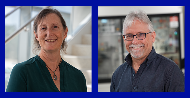
Mary Munson, Craig Peterson named American Association for the Advancement of Science fellows
Read more


It’s not just what we do. It’s how we do it.
Learn in an environment where students drive world-class research and innovation in areas such as biochemistry, biostatistics, cancer biology and neuroscience.
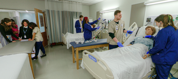
Prepare for a career as a nurse, nurse practitioner or nurse scientist, led by distinguished faculty who promote health equity to improve health and quality of life.
Pursue one of the nation’s best primary care educations, with a focus on patient-centered care in urban and rural communities or an MD/MBA or MD/PhD option.
MassBiologics of UMass Chan Medical School, with locations in Boston and Fall River, is the only nonprofit, FDA-licensed manufacturer of vaccines and biologics in the United States.
ForHealth Consulting is the public service consulting and operations division of UMass Chan Medical School.