

Ancillary Equipment
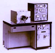
Denton 502 Vacuum Evaporator
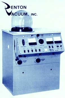 The Denton 502 vacuum evaporation unit is used for vaporizing carbon used to support specimens on electron microscope grids. It is also used to vaporize heavy metals such as platinum, gold, and tungsten used in rotary shadowing visualization of single molecules and molecular assemblies.
Reichert CS Auto freeze-substitution apparatus
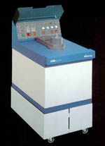 When the ultimate in ultrastructural preservation of tissues or cells is required, with minimal artifacts from specimen preparation, tissues or cells can be ultra-rapidly frozen against a ‘metal mirror’ cooled with liquid helium. Following freezing, specimens are fixed at low temperature (stabilizing cellular components) in a process known as freeze-substitution. They are then embedded and sectioned in the normal way. The facility possesses the capability to carry out both rapid freezing and freeze substitution. The Reichert CS auto allows for precise programming of controlled temperature changes during the cryo-substitution process.
Reichert ultramicrotomes and cryo-attachments
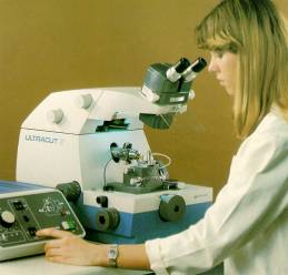
The facility houses two Reichert Ultracut E ultramicrotomes, used for cutting ultrathin sections, on either glass or diamond knives, for transmission electron microscopy. These instruments are the ‘Cadillac’ of the ultramicrotome world. They are extremely well designed, robust and easy to use, and generate superb sections.
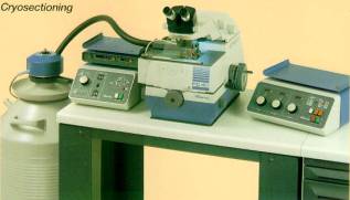
The facility also possesses cryo-attachments for these microtomes, making them capable of cutting ultrathin cryo-sections, which are of prime value in immunolabelling studies when conventional methods must be avoided.
Balzers 401 Freeze-fracture unit
 The facility houses a Balzers 401 freeze-fracture/freeze-etch machine which is used for two main purposes. It provides conventional 'freeze-fracture' images of frozen specimens which reveal the distribution of transmembrane proteins in the plasma and intracellular membranes. When rapidly frozen specimens are 'deep-etched' following fracturing, water is sublimed away from the cytoplasm, exposing the cytoskeleton and organelles, which can be observed in rich three-dimensional detail in the electron microscope.
Canon Flatbed Scanner
The facility possesses a Canon CanoScan 9950F flatbed scanner with transparency adapter for digitizing images directly from electron micrograph negatives. Digitized images can then be manipulated, combined, labeled, etc. in photo-editing programs to produce finished digital files ready for printing or transmission as a digital file.
Darkroom Facilities
The Facility possesses darkrooms for the processing of electron micrograph negatives and for negative printing (2 Durst enlargers and Ilford print processor).