


Apr 1, 2024
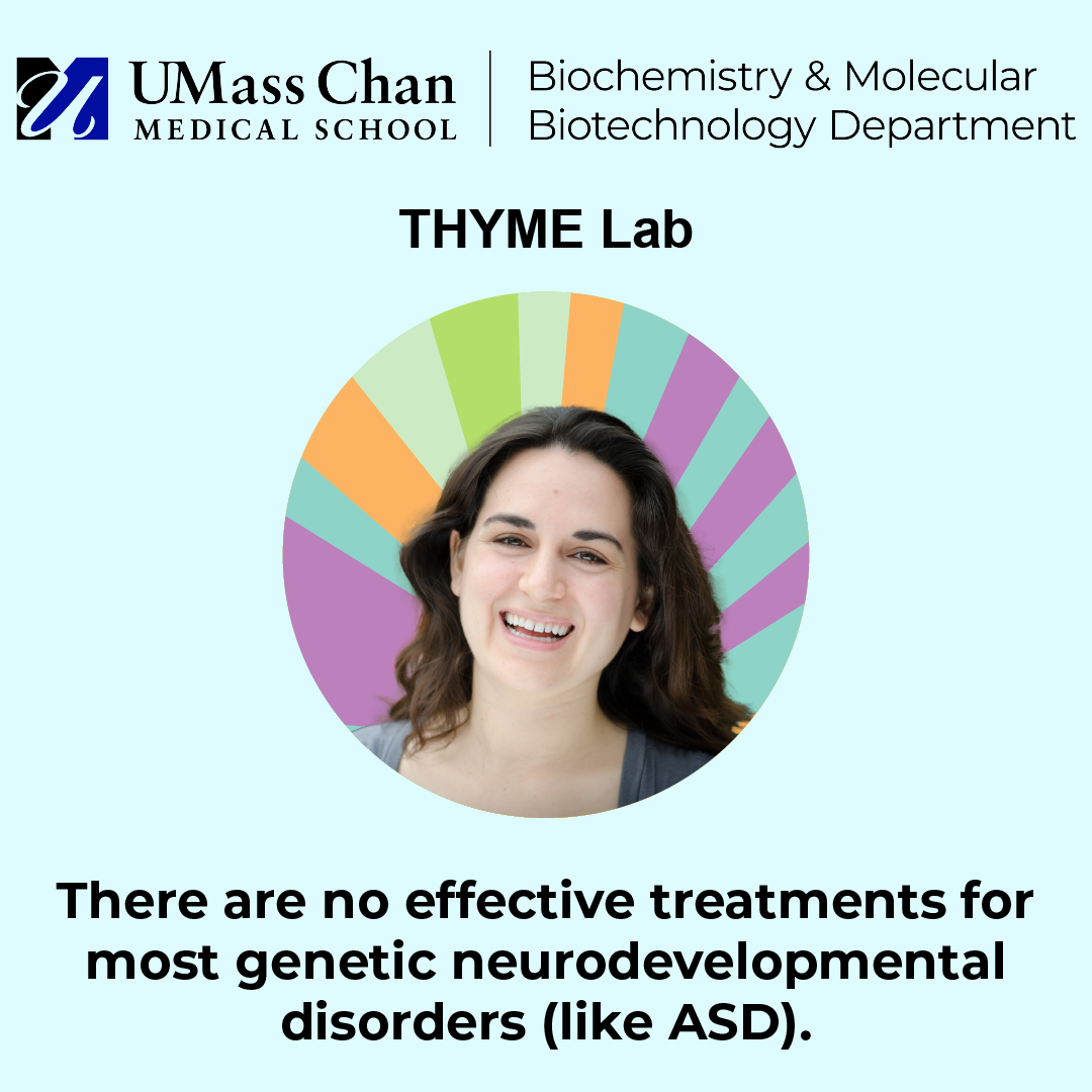
Autism Awareness Day - Featuring the Thyme Lab
Read more

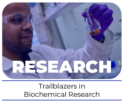
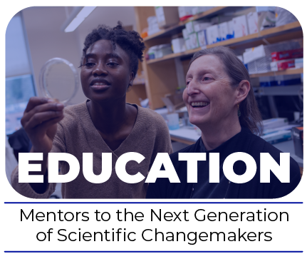
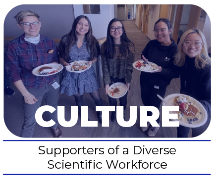
Our mission is to illuminate the molecular mechanisms that govern basic biological processes, with an emphasis on integrative strategies to reveal fundamental mechanistic insights and develop new therapeutic paradigms empowered by a collaborative, diverse & inclusive community.
There are no results for this RSS feed at this time.
Jacob Landeck - Thesis Defense
James E. Ferrell
No RIPS today
Tom Maniatis
RIPS Emma Sedivy
Patriots' Day - closed
No RIPS today
Lindsay Ingerman
RIPS Jaqueline Martinez
Today